XB-IMG-133201
Xenbase Image ID: 133201

|
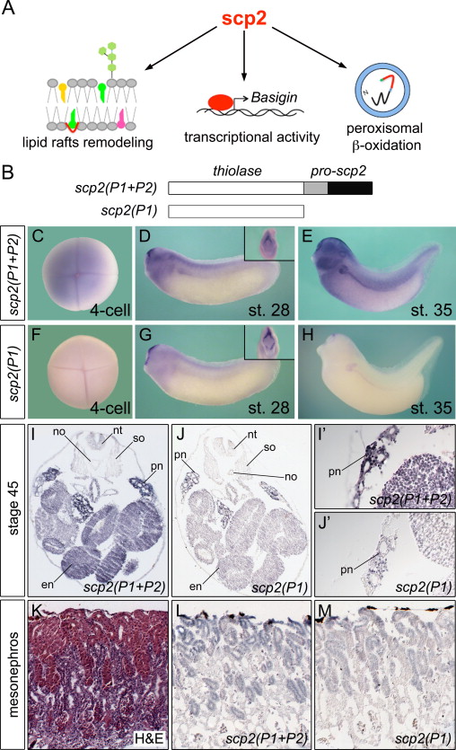
Fig. 2. Expression of Scp2 mRNA during Xenopus Development. (A) Scheme depicting the multiple described activities of scp2 protein. (B) Schematic of the two different antisense probes used. (CâH) Whole mount in situ hybridization of 4-cell stage, stage 28 and stage 35 embryos using the scp2(P1+P2) or the scp2(P1) probe. Inset in (D) and (G) shows a frontal view highlighting the hatching gland expression. (IâJ׳) In situ hybridization on paraplast sections of stage 45 embryos using both scp2 mRNA probes. Panels (I׳) and (J׳) show close-ups of the pronephric kidney area. no, notochord; nt, neural tube; so, somites; pn, pronephros; en, endoderm. (KâM) Hematoxylin and Eosin (H&E) staining (K) and in situ hybridization on paraplast sections using scp2(P1+P2) or scp2(P1) RNA probes (L,M) of an adolescent Xenopus mesonephros. Image published in: Cerqueira DM et al. (2014) Copyright © 2014. Image reproduced with permission of the Publisher, Elsevier B. V.
Image source: Published Larger Image Printer Friendly View |
