XB-IMG-133742
Xenbase Image ID: 133742
|
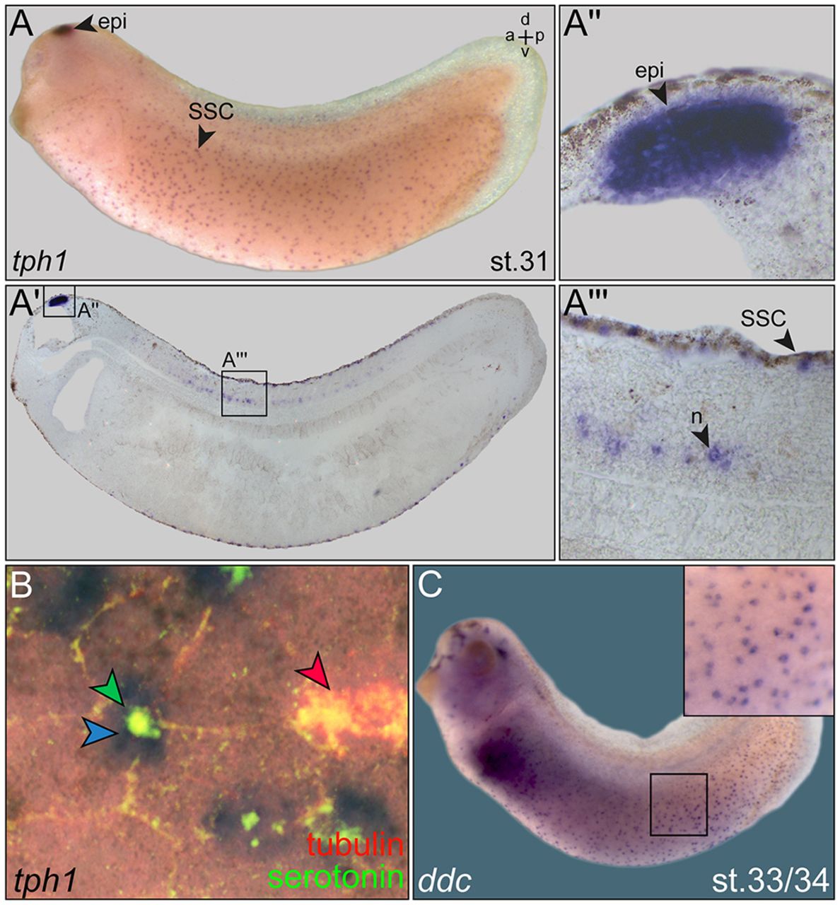
Fig. 2. SSCs express tph1. (A) Expression pattern of tph1 mRNA at stage 31. Signals were detected in the brain and in a punctate pattern in the epidermis. A sagittal histological section (Aâ²) demonstrated tph1expression in the epiphysis (Aâ²) in SSCs and in a subset of neuronal cells (n) in the floor plate of the neural tube (Aâ²â²). (B) SSCs express tph1 (blue arrowhead), as demonstrated by whole-mount in situ hybridization followed by IF for serotonin (green, green arrowhead) and cilia (acetylated-α-tubulin, red, red arrowhead). (C) Punctate expression pattern of aromatic-L-amino-acid decarboxylase (DOPA-decarboxylase; ddc) in the embryonic skin. Inset shows close-up. Embryos are shown in lateral views. a, anterior; d, dorsal; epi, epiphysis; l, left; r, right. Image published in: Walentek P et al. (2014) Copyright © 2014. Image reproduced with permission of the Publisher.
Image source: Published Larger Image Printer Friendly View |