XB-IMG-135204
Xenbase Image ID: 135204

|
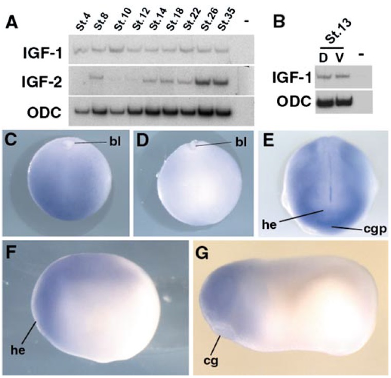
FIG. 1. Expression of IGF signaling components during Xenopus development. (A) RT-PCR analysis of IGF-1 and IGF-2 transcripts throughout development. (B) RT-PCR analysis of IGF-1 expression at early neurula stage (D, dorsal; V, ventral side of the embryo). (CâG) IGF-1R mRNAs detected by in situ hybridization (bl, blastopore; he, head; cgp, cement gland primordium; cg, cement gland). Expression is first detected at the end of the gastrulation (stage 12) in the presumptive dorsal side of the embryo (C) but excluded from the ventral part of the embryo (D). During neurulation, transcripts are present in the head region and in the anterior mesoderm, but are not detectable in the neural tube and the ventral part of the embryo (E, frontal view; F, lateral view). At tailbud stage, transcripts are present in the head and the anterior part of the neural tube (G). Image published in: Richard-Parpaillon L et al. (2002) Copyright © 2002. Image reproduced with permission of the Publisher, Elsevier B. V.
Image source: Published Larger Image Printer Friendly View |
