XB-IMG-135816
Xenbase Image ID: 135816

|
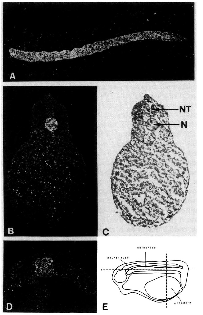
Figure 4. In situ hybridization to sections of Xenopus embryos.
[A] Dark-field illumination shows hybridization of
NC-11 throughout the notochord from the anterior to posterior
region of a stage 25 embryo (see £). [B] Dark-field illumination
shows hybridization to the notochord in a stage 25 embryo. (C)
Bright-field illumination of the section shown in B. (N) Notochord;
(NT) neural tube. (D) Dark-field illumination of a stage
14 embryo shows hybridization to the notochord. (£) Plane of
sections shown in A and B. Although the embryos are at different
stages, the plane of section in D is the same as that in B. Image published in: LaFlamme SE et al. (1988) Copyright © 1988. Image reproduced on with permission of the Publisher, Cold Spring Harbor Laboratory Press. This is an Open Access article distributed under the terms of the Creative Commons Attribution License.
Image source: Published Larger Image Printer Friendly View |
