XB-IMG-1428
Xenbase Image ID: 1428

|
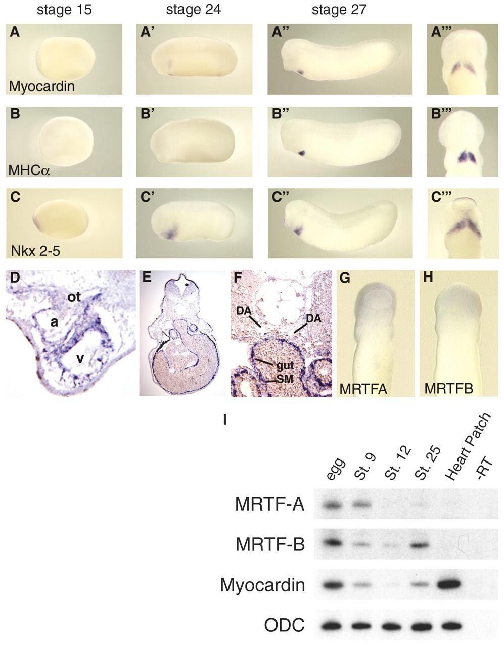
Fig. 2. Developmental expression of Xenopus myocardin and MRTF genes. The expression of Xenopus myocardin (A-A'â) was analyzed by whole-mount in situ hybridization and compared to the expression patterns of the cardiac differentiation marker, MHCα (B-B'â), and the pre-cardiac marker, Nkx2-5 (C-C'â) at the stages indicated. Myocardin expression in the stage 24 embryo is localized to the pre-differentiation cardiac mesoderm in a more restricted domain than Nkx2-5, which is also expressed in the pharyngeal arch region (compare A' with C'). MHCα expression is located in an identical domain to myocardin at stage 27 (compare Aâ with Bâ). A'â, B'â and C'â are ventral views of the stage 27 embryos illustrated. (D) In the heart of a stage 45 embryo myocardin expression is located throughout the myocardial layer of the atrium (a), ventricle (v), and outflow tract (ot). (E) Myocardin is expressed in the visceral smooth muscle in stage 42 embryos. (F) Higher magnification reveals myocardin expression in individual smooth muscle cells adjacent the dorsal aortae and in the smooth muscle layer of the gut. DA, dorsal aorta; SM, smooth muscle. (G, H) In situ hybridization analysis of stage 27 embryos shows that the myocardin-related transcription factors, MRTF-A and MRTF-B, are not expressed in the pre-cardiac mesoderm (ventral views). (I) RT-PCR analysis of myocardin, MRTF-A and MRTF-B expression in early Xenopus embryos and isolated heart patches from stage 28 embryos confirms a lack of MRTF-A and B expression in the pre-cardiac mesoderm. Image published in: Small EM et al. (2005) Copyright © 2005. Image reproduced with permission of the Publisher and the copyright holder. This is an Open Access article distributed under the terms of the Creative Commons Attribution License.
Image source: Published Larger Image Printer Friendly View |
