XB-IMG-145294
Xenbase Image ID: 145294

|
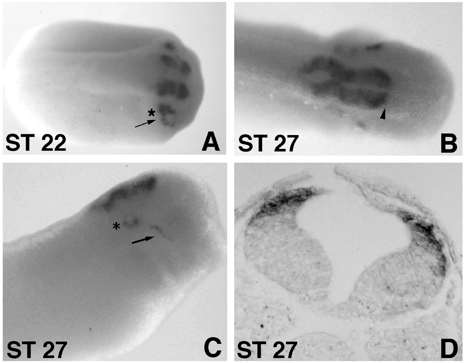
FIG. 2. XATH-1 expression during Xenopus development. Whole mount in situ hybridization analysis was performed during early stages
of Xenopus development. The whole mount in situ hybridization pattern of XATH-1 expression at Stage 22 (A) and Stage 27 (B, C) is
shown. Anterior is to the right. The midbrain/hindbrain boundary is indicated by an arrowhead (B). Expression in the otic vesicle is
indicated by an asterisk and expression in the trigeminal ganglia is indicated by an arrow (A and C). A cross section of a Stage 27 embryo
(D) shows expression within posterior regions of the developing hindbrain. Note that XATH-1-positive cells have exited the ventricular
zone and are now found at the lateral edge of the neural tube. Image published in: Kim P et al. (1997) Copyright © 1997. Image reproduced with permission of the Publisher, Elsevier B. V.
Image source: Published Larger Image Printer Friendly View |
