XB-IMG-145786
Xenbase Image ID: 145786
|
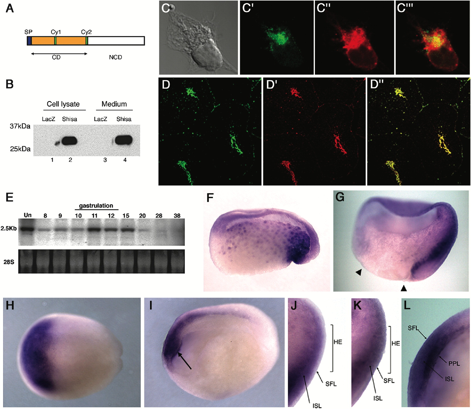
Figure 1.
A Novel Protein Shisa and Its mRNA Expression in the Prospective Head Ectoderm and the Organizer of Xenopus Gastrulae
(A) Schematic structure of Shisa. SP, N-terminal signal peptide; CD, conserved domain; NCD, nonconserved domain; Cy, cysteine-rich domain.
(B) Secretion of Shisa. Western blotting of conditioned medium or cell lysate from HEK 293T cells transfected with shisa-Flag or nuclear-lacZ (control). Molecular weight of secreted Shisa-Flag is 30 kDa.
(CâDâ²) ER localization of Shisa. HEK 293T cells transfected with shisa-Myc (CâCâ´) or a cryosection of the late blastula stage embryo radially injected with 50 pg of shisa-Flag RNA (DâDâ²) were stained for Shisa (green) and an ER marker calreticulin (red).
(E) Developmental Northern blotting of shisa mRNA. Stages of samples are indicated at top. Hybridization with 32P-labeled shisa cDNA is in the middle. Ethidium bromide staining of 28S RNA is at the bottom.
(FâL) Whole-mount in situ hybridization of shisa transcripts. (F, G, J, and K) Sagitally sectioned embryos of stage 10.5 (F) and stage 11.5 (G, J, and K). shisa expression was found in the Spemann organizer of the early gastrulae. In the prospective head ectoderm at mid-gastrula (stage 11.5, according to the developmental time schedule), 55% of the embryos displayed predominant shisa mRNA expression in the internal sensorial layer (ISL in [J]), while 45% had the expression in both superficial layer (SFL) and ISL (K, n = 30). This may be due to slight differences in the developmental stage of each embryo. Arrowheads in (G) indicate blastoderm margin. HE: head ectoderm. (H and L) Dorsal view (H) and sagittally sectioned embryos (L) at the end of gastrulation (stage 13), showing that shisa expression covered the anterior head ectoderm (H) of both ISL and SFL (L). PPL: prechordal plate.
(I) Sagittally sectioned neural plate stage (stage 17) showing downregulated shisa expression in the head ectoderm and persistence in the prechordal plate (indicated by arrow).
Image published in: Yamamoto A et al. (2005) Copyright © 2005. Image reproduced with permission of the Publisher, Elsevier B. V.
Image source: Published Larger Image Printer Friendly View |