XB-IMG-148853
Xenbase Image ID: 148853

|
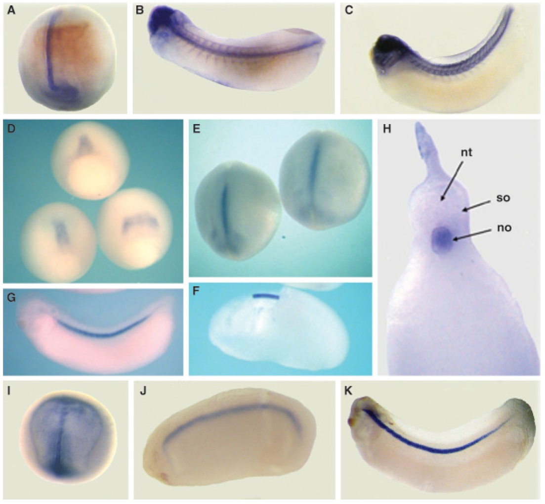
Fig. 3 Spatial expression of Xlox, Xloxl-1, and Xloxl-3. (AâC) Expression
of Xlox was first detected in the notochord at stage 14 (A)
and expanded into the head and somites of tailbud stage embryos
(BâC). (A) Dorsal view of a stage 14 embryo with anterior uppermost.
(B) Dorsal view of a stage 25 embryo with the head on the
left. (C) Lateral view of stage 30 embryo with the head on the left.
(DâH) Expression of Xloxl-1 was first detected in the presumptive
notochord (the dorsal marginal zone) of gastrulae, (D), and continued
to be expressed in the notochord at subsequent stages.
(D) Dorsal-vegetal view of three gastrulae (stages 11â12) with animal
pole uppermost. (E) Dorsal view of two stage 14 embryos with
anterior uppermost. (F) Lateral view of stage 20 embryo with the
head to the left. This embryo has been cracked open to reveal the
intensely stained notochord. (G) Lateral view of a stage 30 embryo
with the head on the left. (H) A cross-section of a St 30 embryo
shows that expression is localized to the notochord (no). The unstained
neural tube (nt) and somites (so) are also labeled. (IâK)
Expression of Xloxl-3 was first detected in the notochord at St 14 (I)
and continued to be expressed in the notochord at subsequent
stages. (I) Dorsal view of stage 14 embryo with head uppermost. (J)
Lateral view of stage 24 embryo with the head to the left. (K)
Lateral view of a stage 28 embryo with the head to the left. Image published in: Geach TJ and Dale L (2005) Copyright © 2005. Image reproduced with permission of the Publisher.
Image source: Published Larger Image Printer Friendly View |
