XB-IMG-1505
Xenbase Image ID: 1505

|
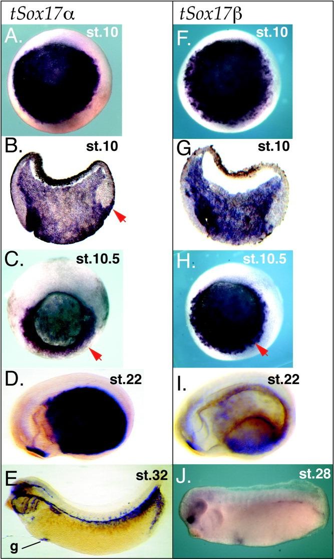
Figure 5. Xenopus tropicalis Sox17 alpha / beta . In situ hybridization analysis of tropicalis embryos with Sox17 alpha (A-E) and Sox17 beta (F-J) antisense probes. Whole-mount (A,C,F,H) and sectioned (B,G) gastrula show that tSox17 alpha and tSox17 beta are identically expressed in throughout the deep and superficial endoderm (red arrows). D: At early tail bud stage (anterior to the left), tSox17 alpha is strongly expressed in the posterior endoderm but absent in the anterior endoderm, except behind the cement gland. I: By comparison at stage 22 (anterior to the left), tSox17 beta transcripts are almost absent from the entire endoderm, except behind the cement gland. E: At stage 32, tSox17 alpha expression persists in the extreme posterior endoderm, the gall bladder (g) precursors and in endothelial cells. J: In late tail bud stages, tSox17 beta is only expressed in a small patch of cells in the head. st, embryonic stage. Image published in: D'Souza A et al. (2003) Copyright © 2003. Image reproduced with permission of the Publisher, John Wiley & Sons.
Image source: Published Larger Image Printer Friendly View |
