XB-IMG-1517
Xenbase Image ID: 1517

|
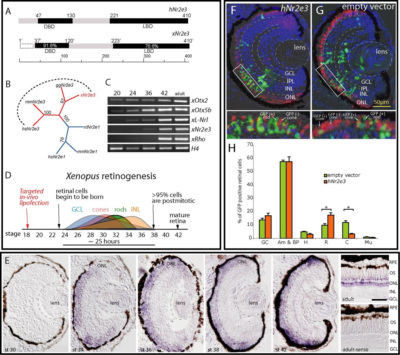
Figure 1. Xenopus Nr2e3 gene is highly conserved with other vertebrates. A: Comparison of human Nr2e3 with Xenopus Nr2e3. The sequence identities of DNA binding domains (DBD) and ligand binding domains (LBD) were revealed by using DNAStar (Stratagene). Numbers in the bars show the percentage of amino acid (AA) sequence identity to hNr2e3. The numbers above the bar indicate the boundaries of different regions based on AA residue position. Xenopus AA residue positions are relative due to the fact that exon 1 is missing. The dashed line represents the unidentified sequence. B: Phylogenetic relationship of Nr2e3 and Nr2e1 family members. Maximum parsimony algorithm with bootstraping was used to determine relationships. Species abbreviations are as follows: hs, Homo sapiens; mm, Mus musculus; xl, Xenopus laevis; gg, Gallus gallus. C: Reverse transcriptase-polymerase chain reaction (RT-PCR) of adult Xenopus heads (stages 20, 24, 36), eyes (stage 42). and retina (adult) RNA using xOtx2, xOtx5b, xL-Nrl, xNr2e3, xRho, and H4 (loading control) primers. D: Timing of Xenopus retinogenesis. E: In situ hybridizations of Xenopus laevis retina at different stages with xNr2e3 probe. Staining can be seen in the outer nuclear layer of the photoreceptors only when antisense probe is used. The negative control (using sense probe) does not produce any staining. Scale bar is 50 mu m. F,G: Embryos at stage 17-18 were lipofected in eye anlagen with either CMV-empty vector (F) or CMV-hNr2e3 together with CMV-GFP (G) and immunostained with cone antibody (anti-calbindin) and DAPI. Cones appear red (untransfected) or yellow (transfected), while rods always appear green (F,G lower panels). The fates of lipofected cells were determined and quantified as the average percentage of each cell type. H: Student's t-test was used to determine significance. Asterisks denote P < 0.05. GC, ganglion; Am&BP, amacrine and bipolar, H, horizontal; R, rod; C, cone; Mu, Müller glia; RPE, retina pigment epithelia; OS, outer segment; ONL, outer nuclear layer; INL inner nuclear layer; IPL inner plexiform layer; GCL, ganglion cell layer. Image published in: McIlvain VA and Knox BE (2007) Copyright © 2007. Image reproduced with permission of the Publisher, John Wiley & Sons.
Image source: Published Larger Image Printer Friendly View |
