XB-IMG-155936
Xenbase Image ID: 155936

|
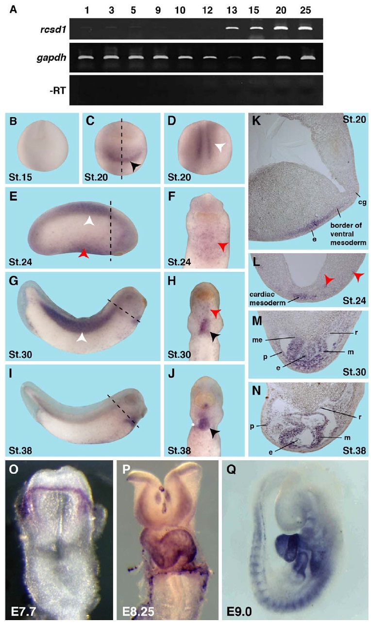
Fig. 2. : Expression of rcsd1 during embryogenesis. A. Temporal expression of rcsd1 at different stages of Xenopus development. Gapdh was used as loading control. Negative controls (-RT) were performed without reverse transcriptase (RT). Rcsd1 is weakly expressed maternally. Zygotic rcsd1 expression starts at stage 13. B-N. Spatial expression of rcsd1 at different developmental stages as indicated in Xenopus. Rcsd1 expression is first detected in the common cardiac progenitor at stage 20 (black arrowhead). Cardiac expression persists until later stages (black arrowheads). In addition, rcsd1 transcripts were detected in the somites (white arrowheads) and myeloid cells (red arrowheads). KN. Cross section through the cardiogenic region of Xenopus embryos as indicated by the black dashed lines in C, E, G and I, respectively. Expression in cardiac tissue is detected at stage 20 (K, sagittal section), 24 (L, transversal section), 30 (M, transversal section)
and 38 (N, transversal section). e, endocardium; m, myocardium; me, mesocardium; p, pericardium; r, pericardial roof. O-Q Expression of Rcsd1 during mouse embryogenesis. Rcsd1 is expressed in the developing murine heart at embryonic stages E7.7 (O), E8.25 (P) and E9.0 (Q). Image published in: Hempel A et al. (2017) Copyright © 2017. Image reproduced with permission of the Publisher, Elsevier B. V.
Image source: Published Larger Image Printer Friendly View |
