XB-IMG-175539
Xenbase Image ID: 175539

|
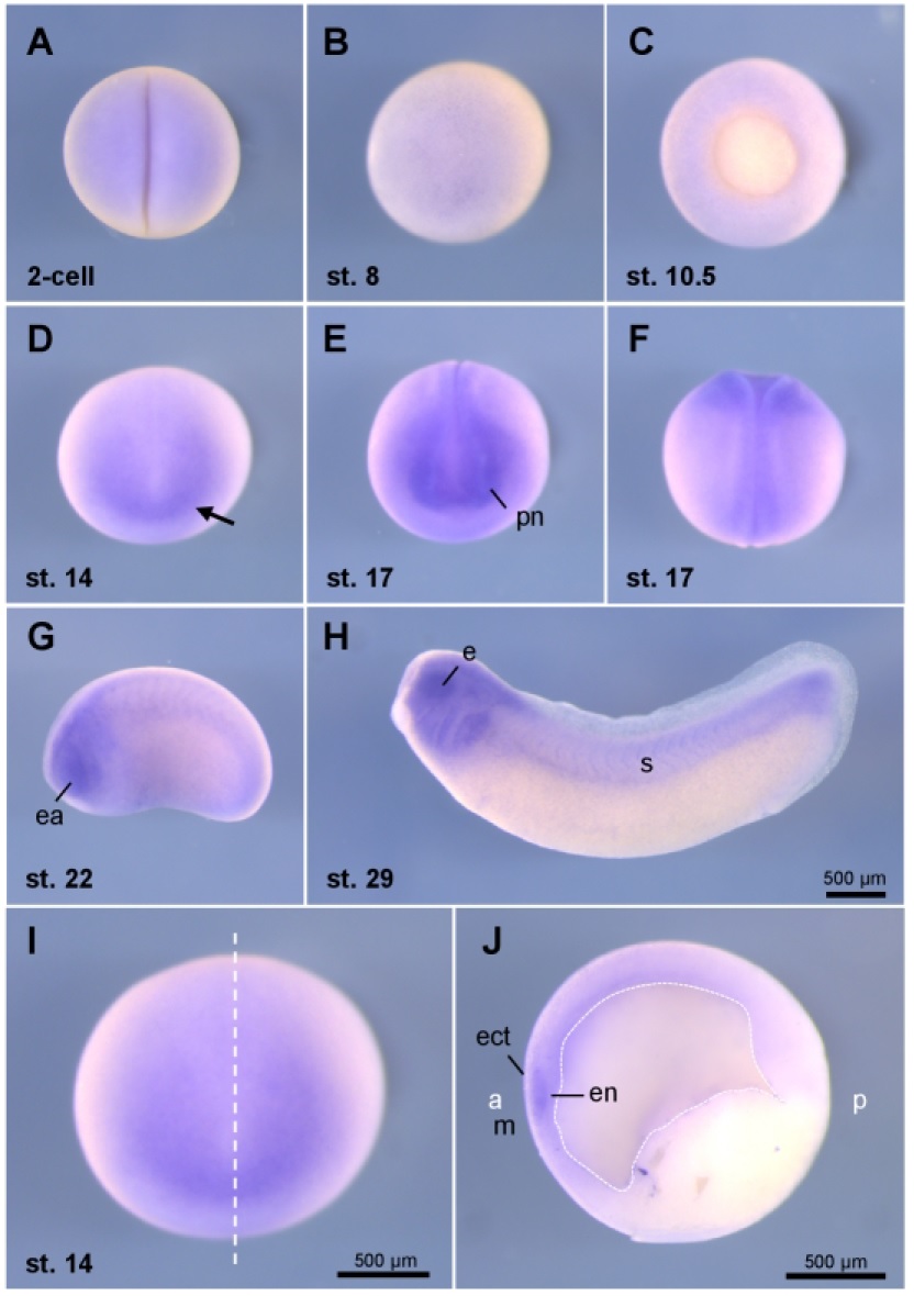
Figure 1: Kmt2d is expressed at early stages of Xenopus development. Kmt2d
expression was analyzed by in situ hybridization. A 2-cell stage embryo. B Embryo at
blastula stage 8. C Embryo at gastrula stage 10.5. D Embryo at neurula stage 14,
anterior view. Arrow marks Kmt2d expression in the anterior region. E Embryo at
stage 17, anterior view. F Same embryo as in E, dorsal view. G Embryo at stage 22,
lateral view. H Embryo at stage 29 lateral view. I Embryo at neurula stage 14, the
dashed line indicates the plane of the sagittal section shown in J. Abbreviations: a
(anterior), e (eye), ea (eye anlage), ect (ectoderm), en (endoderm), m (mesoderm), s
(somites), p (posterior), pn (premigratory neural crest). Image published in: Schwenty-Lara J et al. (2019) Copyright © 2019. Image reproduced with permission of the Publisher, John Wiley & Sons.
Image source: Published Larger Image Printer Friendly View |
