XB-IMG-26235
Xenbase Image ID: 26235

|
||||||||||
|
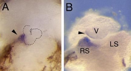
Figure 3. These micrographs, viewed from frontal, show the pericardial cavity, the heart and the cone-shaped accumulation of mesothelial cells (marked by arrows) subsequent to whole-mount in situ hybridization (ISH) with a probe for the proepicardium (PE) marker gene Tbx18 (embryos at stage 41). Tbx18 is strongly expressed in the cone-shaped accumulation of mesothelial cells on the right horn of the sinus venosus but is not expressed on the left sinus horn. Abbreviations as used before. Image published in: Jahr M et al. (2008) Copyright © 2008. Image reproduced with permission of the Publisher, John Wiley & Sons.
Image source: Published Larger Image Printer Friendly View |
