XB-IMG-41229
Xenbase Image ID: 41229

|
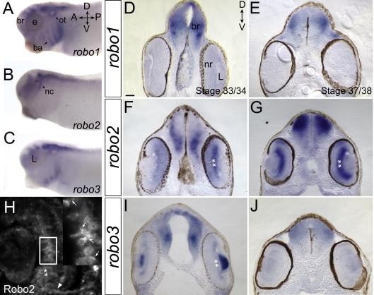
Fig. 2. robo genes are differentially expressed in the developing Xenopus retina. (Aâ G, Iâ J) Xenopus embryos processed for wholemount in situ hybridization for robo1, robo2, or robo3, shown as whole embryos (Aâ C), or in transverse vibratome sections (Dâ G, Iâ J). Robo1 expression is evident in the brain, otic vesicle and branchial arches (A). In stage 33/34 and 37/38 sections, robo1 label is present in the brain, but not in the retina (Dâ E). Robo2 is strongly expressed in migrating neural crest cells (B). Robo2 expression in the brain and eye is already strong at stage 33/34 (F), and becomes even more robust by stage 37/38 (G). Note the strong robo2 label in the GCL (**), with some weaker expression in the other retinal layers (Fâ G). (H) A Robo2 antibody was used to label a transverse cryostat section of a stage 37/38 retina. Robo2 protein is most readily detected in the RGCL (**) and the optic nerve head (arrowhead). The inset in H is an enlargement of the boxed region and shows a few RGCs and their dendrites (arrows) that are expressing Robo2. In wholemounts, robo3 mRNA is detectable in the brain, lens, branchial arches, and migrating cranial neural crest cells (C). A transverse section through a stage 33/34 embryo shows that robo3 mRNA is strongly expressed in the lens, but also weakly in the GCL (**, I). Expression in both locations is lost at stage 37/38 (J). Scale bar in D is 50â î¼m for Dâ G and Iâ J and 25â î¼m for H. A, anterior; br, brain; ba, branchial arches; D, dorsal; e, eye; L, lens; nc, neural crest cells; nr, neural retina; ot, otic vesicle; P, posterior; V, ventral. Image published in: Hocking JC et al. (2010) Copyright © 2010. Image reproduced with permission of the Publisher, Elsevier B. V.
Image source: Published Larger Image Printer Friendly View |
