XB-IMG-41488
Xenbase Image ID: 41488

|
|||||||||||||||||||||||||
|
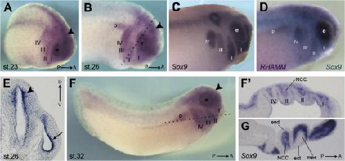
Fig. 3. Spatial expression of RHAMM mRNA during cranial neural crest cells migration. (A) Lateral view of stage 23 embryo showing RHAMM gene expression in the eye field (e), in the neural tube (arrowhead) and in the cranial NCC migrating into the four branchial pouches (I, II, III, IV). (B) At stage 26 RHAMM mRNA is expressed in the eye field (e), in the neural tube (arrowhead), in the NCC migrating into the four branchial pouches (I, II, III, IV) and in the pronephric anlagen (p). (C) The expression of the cranial NCC marker Sox9 is shown for comparison in a stage 26 embryo. (D) Double in situ hybridization showing the overlapping expression of RHAMM (dark blue) with that of the NCC marker Sox9 (light blue) in a stage 26 embryo. (E) Magnification of a coronal vibratome section across the broken line in B highlighting RHAMM expression in the ventricular zone of the neural tube (arrowhead) and in the proliferating region of the optic vesicle (harrow). (F) Lateral view of a stage 32 embryo showing RHAMM transcription in the eye (e), in the neural tube (arrowhead), in the cranial NCC migrated into the four branchial pouches (I, II, III, IV) and in the pronephric anlagen (p). (Fâ²) Vibratome horizontal section across the broken line in C showing RHAMM gene expression in the NCC component of the four branchial arches (I, II, III, IV). (G) The expression of the skeletogenic NCC marker Sox9 is shown for comparison. ect, ectodermal component of the pharyngeal pouches; mes, mesodermal component of the pharyngeal pouches; end, endodermal component of the pharyngeal pouches. Image published in: Casini P et al. (2010) Copyright © 2010. Image reproduced with permission of the Publisher, Elsevier B. V.
Image source: Published Larger Image Printer Friendly View |
