XB-IMG-42724
Xenbase Image ID: 42724

|
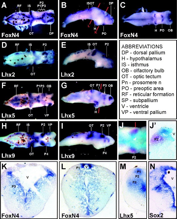
Fig. 3. Expression of X. laevis FoxN4 in the tadpole brain. Visualization of FoxN4 expression in isolated tadpole brains by WISH. Brains were isolated from fixed st 45 tadpoles and subjected to WISH using antisense riboprobes for FoxN4 (AâC), Lhx2 (D,E), Lhx5 (F,G), or Lhx9 (H,I) as indicated. Brain regions are labeled (abbreviations are explained in box). (A,D,F,H) dorsal views; (B,E,G,I) lateral views; (C) ventral views. (J) Simultaneous visualization of FoxN4 (turquoise) and Lhx9 (magenta) expression in the region of prosomere 2 by double in situ hybridization performed using an isolated st 45 tadpole brain. (Jâ²) Section of brain shown in J. (KâM) Sections of isolated tadpole brains subjected to WISH using FoxN4 (K,L) or Lhx5 (M) antisense riboprobes. (N) In situ hybridization performed on a sectioned st 41 tadpole probed with an antisense Sox2 riboprobe. Red lines in B, G, and J indicate planes of section shown in panels Jâ², K, L, and M. Image published in: Kelly LE et al. (2007) Copyright © 2007. Image reproduced with permission of the Publisher, Elsevier B. V.
Image source: Published Larger Image Printer Friendly View |
