XB-IMG-42862
Xenbase Image ID: 42862

|
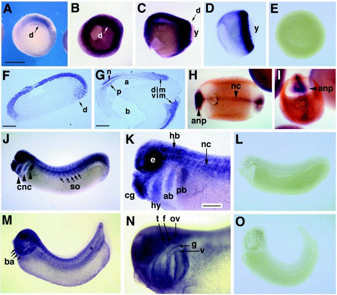
Fig. 4. Localization of rac mRNA by whole-mount in situ hybridization. Xrac mRNA is detected in cells of the marginal zone during early and late gastrulation (A,B, stage 10.5, vegetal view; C, stage 12, lateral view). âdâ indicates the position of the dorsal blastopore lip, and âyâ indicates the yolk plug of the blastopore. The embryo in (A) is uncleared. Xbra expression in an early gastrula-stage embryo indicates the location of presumptive mesodermal cells for comparison (D). Sagittal paraffin sections of hybridized gastrula-stage embryos (F,G). At stage 10.5, rac mRNA is abundant in the presumptive ectoderm and is also detected at elevated levels in cells of the dorsal involuting marginal zone (F). âdâ marks the dorsal blastopore groove. In (G), a sagittal section of the stage 12 embryo shown in whole-mount in (C) reveals rac transcripts specifically in cells of the dorsal (dim) and ventral (vim) involuting mesoderm, the prechordal plate (p), and the sensorial layer of the neuroectoderm (n). The archenteron (a) and blastocoel (b) are indicated. In late neurula-stage embryos, Xrac is expressed in the anterior neural plate (anp) and the notochord (nc) (H,I). At stage 25, Xrac is expressed in the cement gland (cg), the eye (e), the brain (the rhombencephalon (rh) is indicated), the notochord (nc), and somites (so), and is also expressed in the cranial neural crest (cnc), including cells of the hyoid (hy), and anterior (ab) and posterior (pb) branchial streams (J,K). At tailbud stage 33/34, Xrac expression persists in the eye and central nervous system, and is evident in the branchial arches (ba), the otic vesicle (ov), and in the trigeminal (t), facial (f), glossopharyngeal (g), and vagus (v) cranial nerves (M,N). Gastrula-, hatching-, and tailbud-stage embryos were incubated with Xrac sense probe to control for nonspecific hybridization (E,L,O). Scale bar equals 0.5 mm in (AâE,HâJ,L,M,O), and 0.2 mm in (F,G,K,N). Image published in: Lucas JM et al. (2002) Copyright © 2002. Image reproduced with permission of the Publisher, Elsevier B. V.
Image source: Published Larger Image Printer Friendly View |
