XB-IMG-43487
Xenbase Image ID: 43487
|
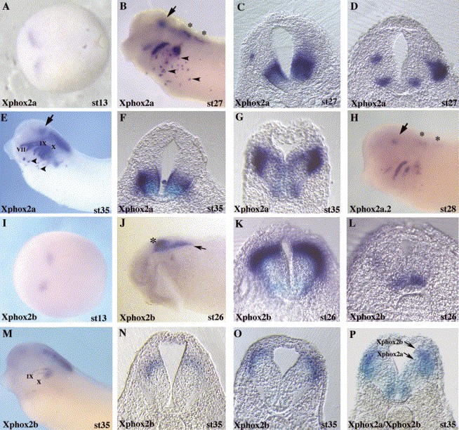
Fig. 2. Characterization of Xphox2a, Xphox2a.2 and Xphox2b expression in the Xenopus embryo. Whole-mount in situ hybridization was used to characterize Xphox2 gene expression during Xenopus development. Xphox2a (AâG) and Xphox2a.2(H) expression in the developing embryo. Similar results were obtained with both full-length and 3â² specific Xphox2a and Xphox2a.2 probes as outlined in the text and Experimental Procedures. Panels (AâG) show staining with full-length Xphox2a probe. Arrow marks ventral midbrain domain of Xphox2a expression at stage 27 (B) and stage 35 (E). This ventral domain is further documented in transverse sections at these stages (C,F). Asterisks mark zones of Xphox2a and Xphox2a.2 expression in the hindbrain (B,H). Transverse sections through posterior hindbrain domains of Xphox2a stained embryos are shown at stage 27 (D) and stage 35 (G). Note that the stage 27 embryo was imbedded asymmetrically and staining in (D) is therefore asymmetric due to the combination of thick sections (30 μm) and discontinuous zones of Xphox2a expression within this region. (IâO) Xphox2b expression in the developing embryo. Asterisk marks anterior hindbrain domain of Xphox2b expression (J) also shown in transverse section at stage 26 (K) and stage 35 (N). Arrow marks posterior hindbrain Xphox2b staining (J) also shown at stage 26 (L) and stage 35 (O). In (P) a cross-section through the hindbrain of an embryo double hybridized with Xphox2a and Xphox2b probes is shown. The Xphox2a domain is blue and the Xphox2b domain is magenta/purple. Image published in: Talikka M et al. (2004) Copyright © 2004. Image reproduced with permission of the Publisher, Elsevier B. V.
Image source: Published Larger Image Printer Friendly View |