XB-IMG-43531
Xenbase Image ID: 43531
|
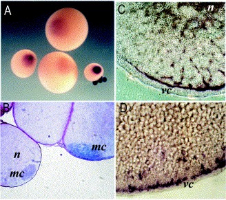
Fig. 3. Localization of Germes mRNA during oogenesis analyzed by in situ hybridization. (A) Whole-mount of stages IâVI oocytes, vegetal view. In the smallest oocytes (stage II), the entire cytoplasm is heavily stained. (B) Epon section through stage II oocytes reveals concentration of Germes message in mitochondrial cloud. Oocyte on the right illustrates the commencement of the accumulation of the message in the vegetal cortex. (C) In situ hybridization for Germes on paraffin sections of stage III oocyte. The mRNA anchors on the vegetal cortex, forming the continuous layer. (D) In situ hybridization on paraffin sections of stage VI oocytes. The Germes transcript forms a granular pattern within the vegetal cortex. mc, mitochondrial cloud; n, nucleus; vc, vegetal cortex. Image published in: Berekelya LA et al. (2003) Copyright © 2003. Image reproduced with permission of the Publisher, Elsevier B. V.
Image source: Published Larger Image Printer Friendly View |