XB-IMG-44507
Xenbase Image ID: 44507
|
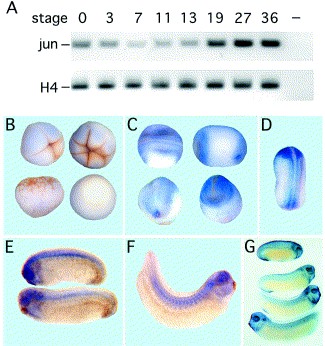
Fig. 2. Xenopus c-Jun expression during embryogenesis. (A) Temporal expression of c-Jun was analyzed with total RNA from embryos of different developmental stages (Nieuwkoop and Faber, 1975) by RT-PCR using oligonucleotides 5â²-GGC AGA AAG GAA GCG TAT GAG-3â² as forward and 5â²-CTG CTG TGT TAA CAT TAG CTC AC-3â² as reverse primer, repectively. Histone H4 was used as internal control. The last lane refers to a control assay lacking RNA. Spatial expression of Xenopus c-Jun was analyzed by whole-mount in situ hybridizations. (B) Four-cell, eight-cell, morula and gastrula stage embryos. Note the weak staining being most probably due to maternal transcripts. (C) Neurula stage embryos from dorsal and posterior (left) or from ventral and anterior (right). (D) Stage 22. c-Jun expression is observed at the posterior and lateral region at the lateral border of the neural plate but not within the dorsal midline. Anterior staining is within the neural plate. (E) Stages 25 and 27. (F) Stage 31/32. c-Jun transcripts are found within trunk neural crest cells, otic vesicle, the eye and head mesenchyme. (G) BMP-4 whole-mount in situ hybridization of embryos at later developmental stages. Note the similarities to the c-Jun expression pattern. Image published in: Knöchel S et al. (2000) Copyright © 2000. Image reproduced with permission of the Publisher, Elsevier B. V.
Image source: Published Larger Image Printer Friendly View |