XB-IMG-44569
Xenbase Image ID: 44569

|
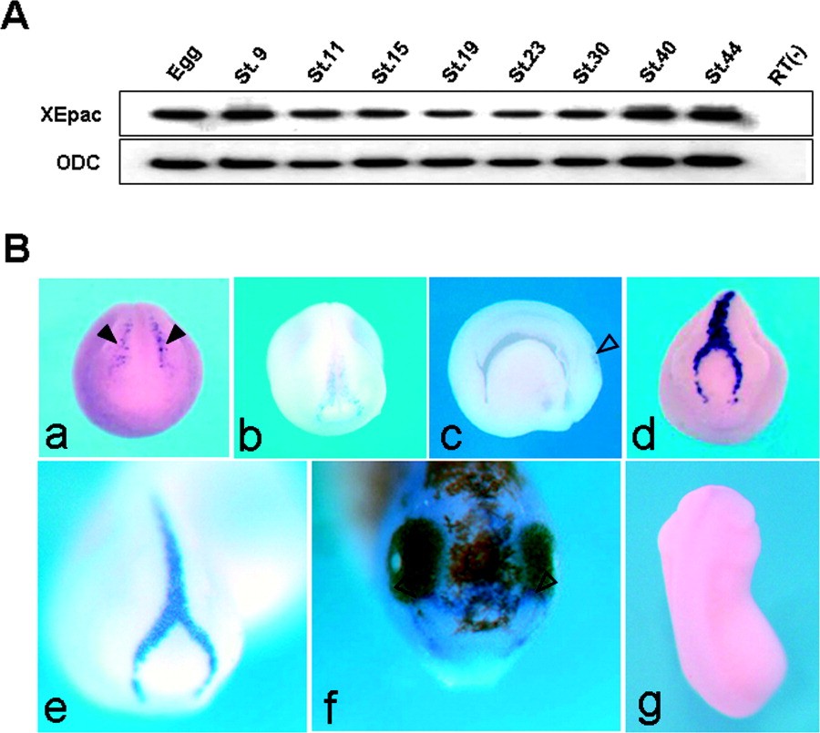
Figure 2. Temporal and spatial expression patterns of XEpac. A: Temporal expression patterns. XEpac was expressed both maternally and zygotically. RT(-), lacking reverse transcriptase. B: Spatial expression patterns. XEpac is restricted exclusively within the developing hatching gland, which forms a characteristic inverted Y type and functions as secreting hatching enzyme. It first appeared in the early neurula and persisted as long as the hatching gland existed (a-f, black and open arrowheads); no positive signals were detected with XEpac sense probe (g). a is early neurula stage; b and c are late neurula and its sagittal section, respectively; d, e, and g are tail bud stage; f is tadpole stage. c is a lateral view, g dorsal; others are anterior or dorsoanterior views. Image published in: Lee SJ and Han JK (2005) Copyright © 2005. Image reproduced with permission of the Publisher, John Wiley & Sons.
Image source: Published Larger Image Printer Friendly View |
