XB-IMG-45954
Xenbase Image ID: 45954

|
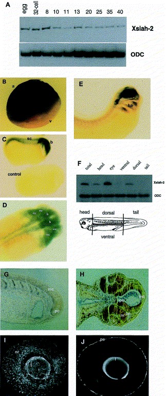
Fig. 3. Temporal and spatial expression pattern of Xsiah-2 during development. (A) RT-PCR detecting Xsiah-2 and ODC transcripts in total RNA of relevant embryonic stages (Nieuwkoop and Faber, 1975). (BâE) In situ hybridization using a digoxygenin labelled Xsiah-2 antisense probe. The Xsiah-2 transcripts were visualized with a phosphatase-coupled α-digoxygenin antibody (BâD,G,H) or with an FITC-coupled α-digoxygenin antibody (I,J). Hybridization results of a fertilized egg (B), a lateral view of the late neurula stage 22 (C), a top view of the head region of a tailbud stage 28 (D) and lateral view of swimming larvae stage 34 (E), the anterior part to the right. The control embryo in (C) (stage 22) was hybridized with a sense probe. (F) RT-PCR to detect Xsiah-2 and ODC transcripts in a dissected stage 39 embryo. (G,H) Sections of a stage 26 (G) and a stage 32 (H) embryo after in situ hybridization with Xsiah-2 antisense RNA. (I,J) Laser scan images of an eye derived from a stage 38 embryo specifically stained for Xsiah-2 transcripts. Staining of the lens is due to autofluorescence. a, animal; v, ventral; sc, spinal cord; b, brain; ev, eye vesicles; nc, neural crest cells; ba, branchial arches; pc, prosencephalon; mc, mesencephalon; nr, neural retina; pe, pigmented epithelium; nr, neural retina; l, lens. Image published in: Bogdan S et al. (2001) Copyright © 2001. Image reproduced with permission of the Publisher, Elsevier B. V.
Image source: Published Larger Image Printer Friendly View |
