XB-IMG-49673
Xenbase Image ID: 49673
|
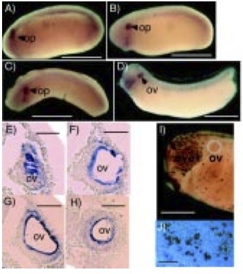
Fig. 3. Expression of xPAK1 in the otic placode and vesicle. Lateral views of whole-mount in situ hybridization of xPAK1 transcripts in X. laevis embryos at (A) stage 23/24, (B) stage 25, (C) stage 26, dorso-lateral view, (D) stage 29. (G-H) Sequential sections of a xPAK1 hybridized stage 29/30. (I) Whole-mount TUNEL staining [(Hensey and Gautier, 1998) as modified by Poitras et al. manuscript submitted] of anterior region of a stage 27 embryo. (J) Magnified view of TUNEL staining over the eye of a stage 27 embryo showing colocalization of stain with cell nuclei, as revealed by Hoechst 33258 fluorescence. Op, otic placode; ov, otic vesicle. Bar in A to D, 1 mm; in E to H, 50 μm; in I 0.5 mm; and in J 100 μm. Image published in: Islam N et al. (2000) Copyright © 2000. Image reproduced with permission of the Publisher.
Image source: Published Larger Image Printer Friendly View |