XB-IMG-49768
Xenbase Image ID: 49768

|
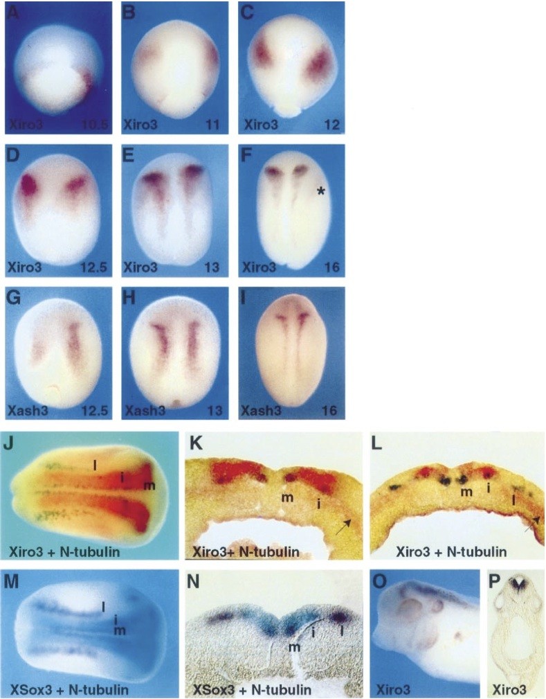
Fig. 2. Xiro3 expression during early embryogenesis. (A) Embryos at the indicated stages are analysed by in situ hybridization with probes for Xiro3 (A) and XASH3 (G). Note that Xiro3 expression within the prospective neural plate is first detected at stage 11 in the form of two wide symmetrical patches (B) and that later Xiro3 and XASH3 are similarly expressed in the neural plate in the form of two symmetrical longitudinal stripes and two transverse anterior bands. In addition, expression of Xiro3 also occurs in the ectoderm lateral to the anterior neural plate (asterisk). (J) Double in situ hybridization of Xiro3 (red) and N-tubulin (purple) in a stage 15 embryo (m, medial; i, intermediate; l, lateral). (K) Cross-sections of a stage 15 Xiro3/N-tubulin-stained embryo showing that Xiro3 is expressed in regions corresponding to the medial and intermediate stripes of N-tubulin and that it is detected in cells located more superficially than those expressing N-tubulin. (L) Cross-section of a stage 15 Xiro3/N-tubulin- stained embryo at a more posterior position. Note that Xiro3 expression appears in both the superficial and deep layer of the neurectoderm. Xiro3 is also expressed in anterior lateral mesoderm (arrowheads). (M) Double in situ hybridization of XSox3 (blue) and N-tubulin (purple). (N) Cross-section of the XSox3/N-tubulin-stained embryo shown in (M). Note that XSox3 is expressed like Xiro3 in the posterior neural plate in superficial layers of the neurectoderm in between the intermediate and medial N-tubulin stripes. (O) Lateral view of the head of a tadpole embryo. (P) Transverse section at the level of the otic vesicle. All embryos are shown in a dorsal view, with anterior to the top, except in (J), (M) and (O). Image published in: Bellefroid EJ et al. (1998) Copyright © 1998. Image reproduced with permission of the Publisher, John Wiley & Sons.
Image source: Published Larger Image Printer Friendly View |
