XB-IMG-74571
Xenbase Image ID: 74571
|
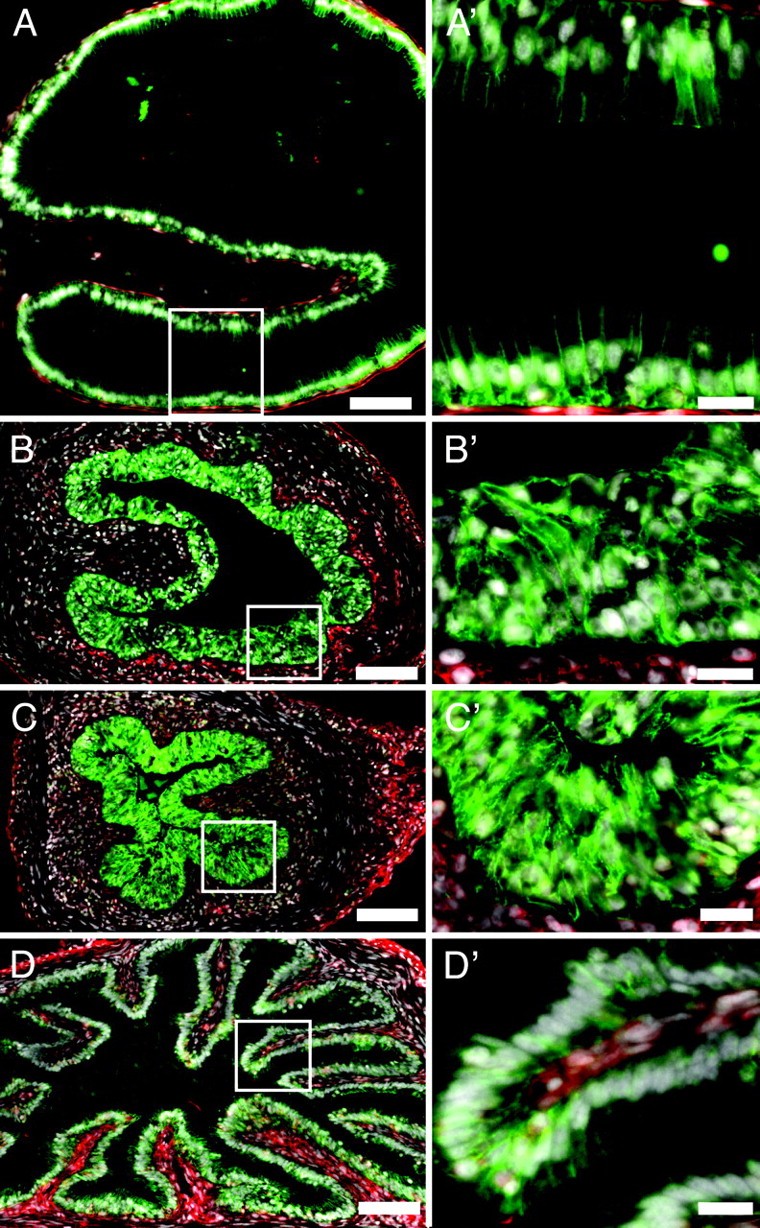
Fig. 2. The intestinal epithelium thickens during metamorphic climax. Cross sections of the duodenum at different developmental stages are labeled with antibodies against proteins that are localized in the epithelium (e-cadherin, green) and mesenchyme (fibronectin, red). Nuclei are labeled with DAPI (white). (A and Aâ²) Before metamorphosis (NF 54), the epithelium is only one to two cell layers thick. During metamorphic climax (B and Bâ²,NF62; C and Câ², NF 63), the epithelial layer becomes five to eight cell layers thick. (D and Dâ²)At the end of climax (NF 66), the epithelium returns to one to two cell layers thick. [Scale bars: 100 μm (AâD) and 20 μm (Aâ²âDâ²).] The boxed regions in AâD correspond with the magnified regions shown in Aâ²âDâ². Image published in: Schreiber AM et al. (2005) Copyright © 2005. Image reproduced with permission of the Publisher.
Image source: Published Larger Image Printer Friendly View |