Click here to close Hello! We notice that you are using Internet Explorer, which is not supported by Xenbase and may cause the site to display incorrectly. We suggest using a current version of Chrome, FireFox, or Safari.
XB-IMG-74580

Xenbase Image ID: 74580
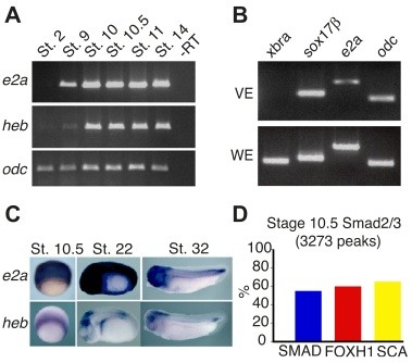

Figure 4. e2a and heb are expressed during early development in X. tropicalis. (A) X. tropicalis embryos were analyzed by RTCR for expression of heb and e2a, from blastula stage through early neurula. Ornithine decarboxylase (odc) was used as a loading control. (B) Vegetal endoderm explants of stage 10.5 X. tropicalis express e2a at low levels, but not the mesoderm- specific gene xbra. (C) X. tropicalis embryos were analyzed by in situ hybridization for expression of e2a and heb at early gastrula stage (stage 10.5), at early tailbud stage (stage 22), and at late tailbud stage (32). (D) Bar graphs show the percentage of each motif present in Smad2/3 target regions during gastrulation in X. tropicalis as determined by ChIP-seq.

Image published in: Yoon SJ et al. (2011)

Copyright © 2011. Image reproduced on with permission of the Publisher, Cold Spring Harbor Laboratory Press. This is an Open Access article distributed under the terms of the Creative Commons Attribution License.

GeneSynonymsSpeciesStage(s)Tissue
tcf3bhlhb21, E12, e2a, itf1, tcf-3, tcfe2a, vdir, XE12, xtcf-3, xtcf3X. tropicalisThroughout NF stage 10.5mesoderm
ectoderm
tcf12HEB, XE1.1X. tropicalisThroughout NF stage 10.5mesoderm
involuting marginal zone
marginal zone
tcf3bhlhb21, E12, e2a, itf1, tcf-3, tcfe2a, vdir, XE12, xtcf-3, xtcf3X. tropicalisThroughout NF stage 22whole organism
tcf12HEB, XE1.1X. tropicalisThroughout NF stage 22lateral plate mesoderm
optic vesicle
hindbrain
presomitic mesoderm
tcf3bhlhb21, E12, e2a, itf1, tcf-3, tcfe2a, vdir, XE12, xtcf-3, xtcf3X. tropicalisThroughout NF stage 32head region
mesoderm
paraxial mesoderm
lateral plate mesoderm
chordoneural hinge
hindbrain
neuroectoderm
pharyngeal arch
pronephric duct
tcf12HEB, XE1.1X. tropicalisThroughout NF stage 32optic vesicle
pharyngeal arch
branchial arch
hindbrain
presomitic mesoderm

Image source: Published

Larger Image
Printer Friendly View

Return to previous page