XB-IMG-76165
Xenbase Image ID: 76165
|
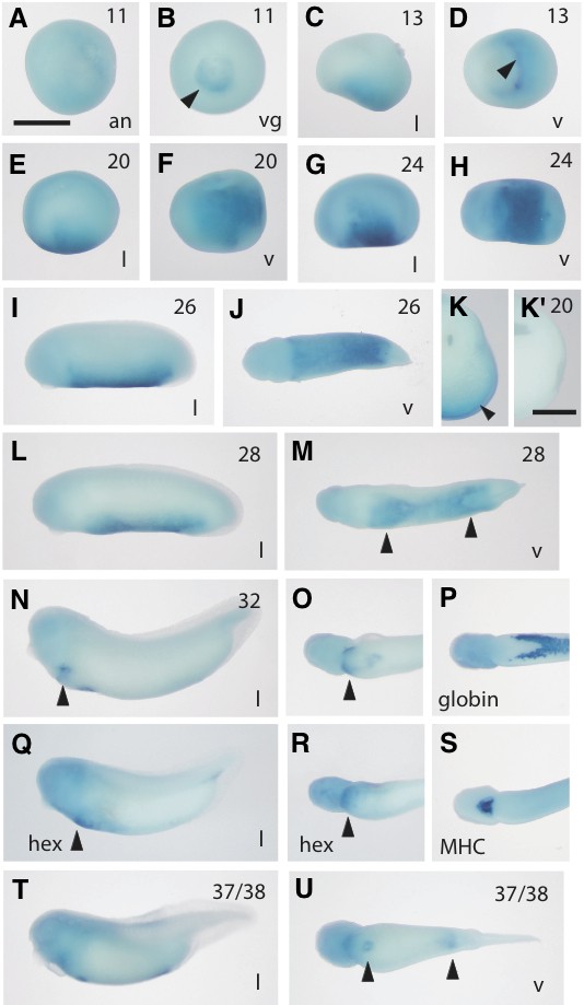
Fig. 3 (Right). Whole-mount in situ hybridization analysis of val in developing embryos. A very faint expression of val message was first detected in the yolk plug (presumptive endoderm) at the mid-gastrula stage (st. 11) (A,B). Expression was gradually accumulated in the posterior ventral part of the neurula embryo (st. 13-20) (C-F) and peaked at the early tailbud stage (st. 24) (G,H). Dissection of the stained embryo (st. 20) showed that val was expressed in all three germ layers (K). Kshows a control dissected embryo hybridized with the sense probe. During the tailbud stages, the expression of val extended toward anterior and posterior directions (st. 26-28) (I,J,L,M) and two domains of expression areas were separated each other (arrowheads in M). Positive areas of val (N,O), α-globin (P), hex (Q,R) and myosin heavy chain (MHC) (S) at the late tailbud stage (st. 32) were compared each other, suggesting that val-positive area overlapped with hex-positive area (a region of the liver rudiment as indicated by arrowheads in N, O, Q and R). Positive messages were visible in the two spots of anterior and posterior regions (arrowheads in U) in the swimming tadpoles (st. 37/38) (T,U). Scale bars in A and Kshow 1 mm and 500 μm, respectively. Image published in: Saito Y et al. (2010) Copyright © 2010. Image reproduced with permission of the Publisher, University of the Basque Country Press.
Image source: Published Larger Image Printer Friendly View |