XB-IMG-76175
Xenbase Image ID: 76175

|
||||||||||||||||||||||||||||||||||||||||||||||||||||||||||||
|
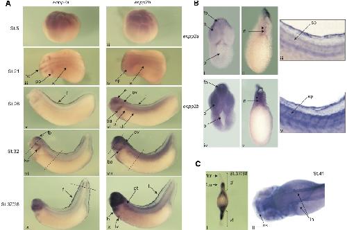
Fig. 7. Comparison of the spatial expression profile of enpp2a [enpp2.S] and enpp2b [enpp2.L] genes during development. (A) Whole-mount in situ hybridisation with an enpp2a and enpp2b DIG labelled anti-sense RNA probe was performed on embryos from stages 5-41. Animal view at stage 5 (i, ii). Lateral view at stage 21 (iii, iv), stage 26 (v, vi), stage 32 (vii, viii) and stage 37/38 (ix, x). Dorsal is up and anterior is left. The dotted line through the fins (ix) corresponds to plane of section in Ci. (B) Whole-mount in situ hybridisation with an enpp2a and enpp2b DIG labelled antisense RNA probe on stage 32 embryos. Transverse section through the head (i, iv) and trunk (ii, v) levels. Lateral view of the trunk region (iii, vi). Arrowheads indicate the staining in the spinal cord. (C) Whole-mount in situ hybridisation with an enpp2a DIG labelled antisense RNA probe. Transverse section through the fin at stage 37/38 (i). Dorsal view of a stage 41 cleared embryo (ii). ba, branchial arches; df, dorsal fin; dt, distal tubules; e, eye; f, fin; fb, forebrain; fcr, fin crest; fco, fin core; h, heart; lh, lymphatic heart; lv, liver diverticulum; n, notochord; nc, neural crest; np, neural plate; ns, nasal placode; ov, otic vesicle; p, pharynx; pt, pronephric tubules; s, somites; sp, spinal cord; vf, ventral fin. Image published in: Massé K et al. (2010) Copyright © 2010. Image reproduced with permission of the Publisher, University of the Basque Country Press.
Image source: Published Larger Image Printer Friendly View |
