XB-IMG-77252
Xenbase Image ID: 77252
|
||||||||||||||||||||
|
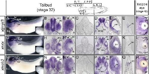
Fig. 4. Expression of xtjmjd3, xtutx and xtezh2 in tailbud embryos and tadpoles. (A,G,M) Lateral views of tailbud embryos subjected to whole-mount in situ hybridiza- tion. Insets show embryos hybridized with a sense probe for xtjmjd3, xtutx or xtezh2. (B-E,H-K,N-Q) Transverse sections sub- jected to in situ hybridization. The planes of the sections are illustrated in the top white box. (BHN High magnification views of eyes in the transverse sections shown in B, H, and N. Arrows indicate the outer layer of the optic cup. (C,I,O) Transverse sections hybridized with a sense probe for xtjmjd3, xtutx or xtezh2. (DJP High magnification views of otic placodes in the transverse sections shown in D, J and P. (F,L,R) Transverse sections of tadpole eyes subjected to in situ hybridization. br, brain;
ov, otic vesicle; ba, branchial arch; mc, mesencephalon; dc, diencephalon; oc, optic cup; le, lens; mnc, migrating neural crest; rc, rhombencephalon; nc, notochord; sc, spinal cord; gcl, ganglion cell layer; inl, inner nuclear layer; prl, photoreceptor layer. Image published in: Kawaguchi A et al. (2012) Copyright © 2012. Image reproduced with permission of the Publisher, University of the Basque Country Press.
Image source: Published Larger Image Printer Friendly View |