XB-IMG-79141
Xenbase Image ID: 79141
|
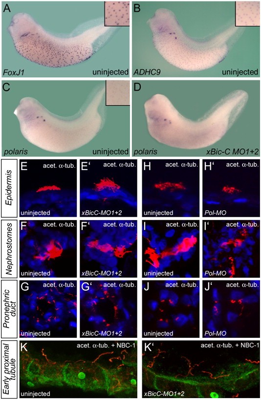
Fig. 6. Bicaudal-C and cilia formation. (AâD) Whole mount in situ hybridization of uninjected (AâC) or xBic-C-MO1 + 2-injected Xenopus embryos (D) at stage 36 using (A) FoxJ1, (B) Axonemal Dynein Heavy Chain 9 (ADHC9) and (C, D) polaris. (EâJâ²) Immunocytochemistry using anti-acetylated α-tubulin antibody (red) on uninjected control, xBic-C-MO1 + 2 and xPol-MO-injected embryos at stage 42. (E, Eâ², H, Hâ²) Ciliated cells of the epidermis; (F, Fâ², I, Iâ²) cilia of the nephrostomes; (G, Gâ², J, Jâ²) individual primary cilia present on the cells of the pronephric duct. (K, Kâ²) Immunocytochemistry using anti-acetylated α-tubulin (red) and anti-NBC1 (green) antibodies on uninjected control and xBic-C-MO1 + 2-injected embryos at stage 42. Images were obtained by confocal microscopy; nuclei were counterstained with DAPI (blue). Image published in: Tran U et al. (2007) Copyright © 2007. Image reproduced with permission of the Publisher, Elsevier B. V.
Image source: Published Larger Image Printer Friendly View |