XB-IMG-79641
Xenbase Image ID: 79641
|
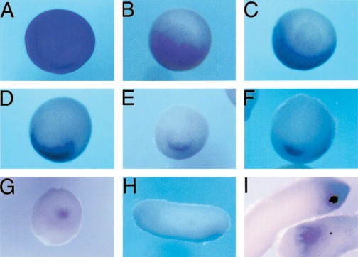
Fig. 3. Xvex-1 is expressed as a ventral gene. Whole mount in situ hybridization analysis was performed to determine the spatial pattern of Xvex-1 expression during early embryogenesis. (A) At stage 10 Xvex-1 transcripts can be observed throughout the embryo (animal view). (B) At late stage 10.5 a wave of repression can be observed spreading from dorsal to ventral on the vegetal side including the marginal zone and the yolk-filled endodermal cells. (C) By stage 11.5 the Xvex-1 transcripts are localized to the ventral marginal zone. (D) The ventral localization of the Xvex-1 continues at stage 12.5 but the gap between the expression domain and the blastopore disappears. (E) The onset of neurulation (stage 13) marks a continued lateral restriction in the Xvex-1 pattern of expression. (F) At stage 14 the process of restriction in the lateral extent of Xvex-1 continues. (G) By stage 16 the Xvex-1 expression domain acquires its minimal size on the ventral-most region of the closed blastopore. By stage 26/27 expression of Xvex-1 remains in the same position on the now ventrally localized proctodeum, (H) lateral view, (I) ventral views. Panels B and C vegetal view, DâG posterior view. Panels (AâH), dorsal is to the top.
Image published in: Shapira E et al. (1999) Copyright © 1999. Image reproduced with permission of the Publisher, Elsevier B. V.
Image source: Published Larger Image Printer Friendly View |