XB-IMG-80321
Xenbase Image ID: 80321
|
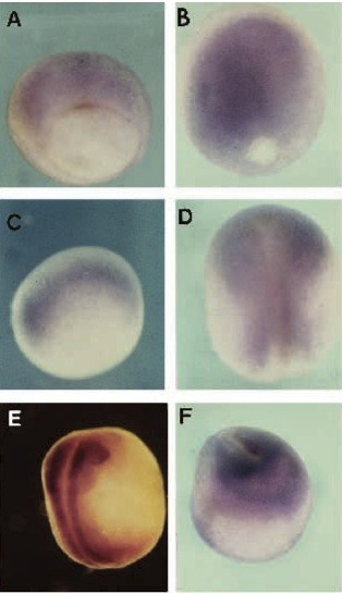
Fig. 3. Expression pattern of XeIF4AII by whole mount in situ hybridization. The eIF4AII-specific probe was derived from the 3â² UTR. (A) Stage 11.5, dorsal aspect; (B) stage 12, dorsal aspect; (C) stage 12, lateral aspect (anterior to the right); (D) stage 16, dorsal aspect; (E) stage 20, embryos cleared; (F) stage 20, anterior end with staining in the prospective cement gland. Image published in: Morgan R and Sargent MG (1997) Copyright © 1997. Image reproduced with permission of the Publisher and the copyright holder. This is an Open Access article distributed under the terms of the Creative Commons Attribution License.
Image source: Published Larger Image Printer Friendly View |