XB-IMG-82019
Xenbase Image ID: 82019

|
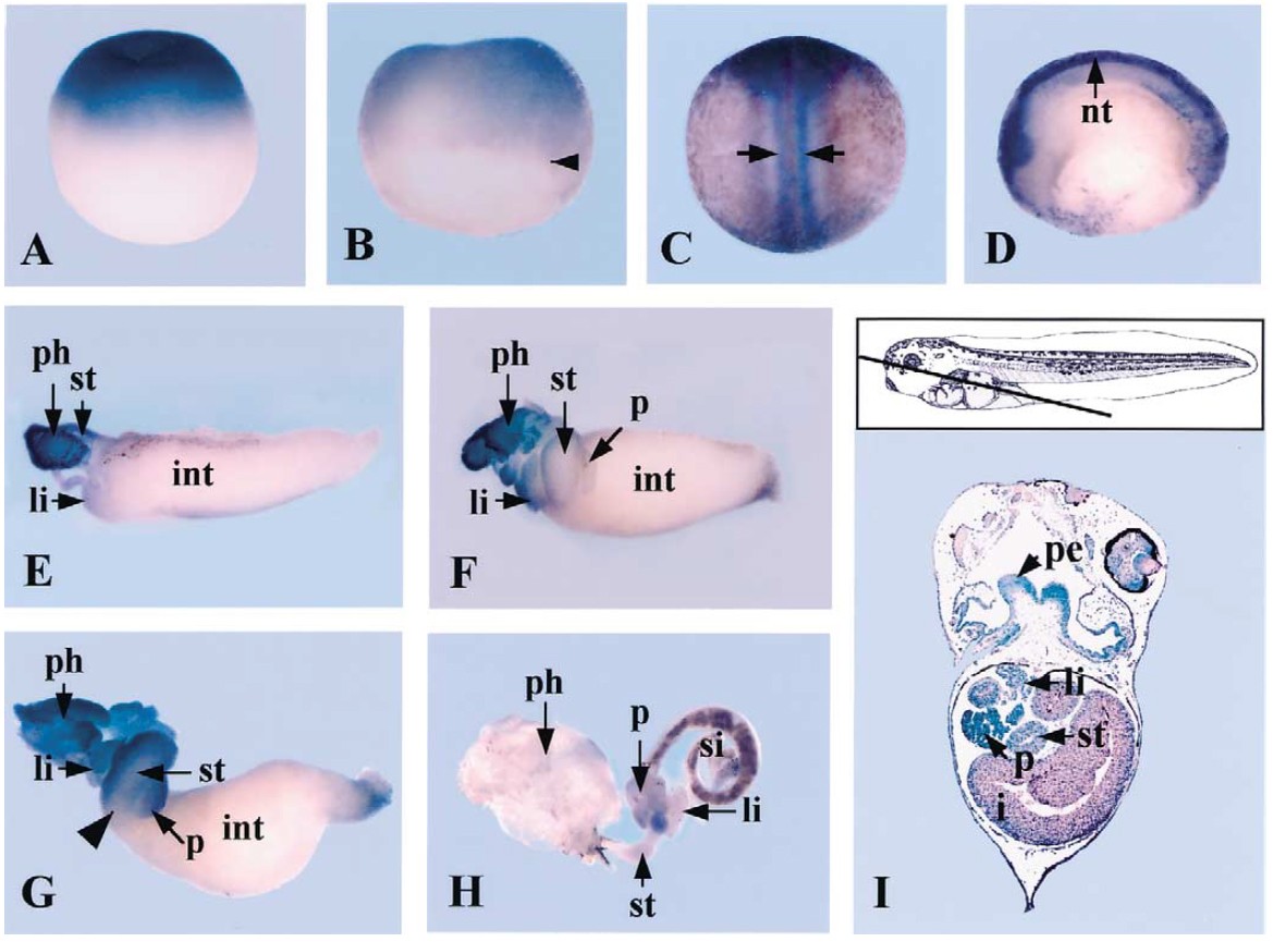
Fig. 3. Localization of Xamp transcripts in whole embryos and isolated guts. (A) The lateral view of a fertilized egg showing that maternal transcripts of Xamp
are localized in the animal region of egg. (B) The dorsolateral view of an early gastrula embryo (stage 10). Xamp mRNA is restricted to the dorsal ectoderm.
Arrowhead indicates the dorsal lip of blastopore. (C) The dorsal view of a neurula embryo (stage 18). Xamp is expressed in the prospective sensory neuron area
of the neural tube (arrow). (D) A sagittal section of stage 18/19 embryo showing Xamp expression in the most dorsal region of neural tube (nt). (E) Isolated
whole guts. (E) Stage 36 embryonic gut shows Xamp localization in the developing pharynx (ph). Anterior is to the left. (F) Left side view of Xamp expression
in the stage 38/39 gut. Xamp is expressed in pharynx (ph) and liver bud (li) of foregut. (G) Ventrolateral view of stage 41 gut. Xamp is expressed in the pharynx
(ph) and foregut including liver (li), pancreas (p), and stomach (st). The posterior expression boundary of Xamp expands to the transitional zone/intestine
boundary (arrowhead). (H) Fully differentiated stage 49 gut. No positive signaling is detected at this stage. (I) A frontal section of the stage 42 tadpole shows
that Xamp expression is detected in the endoderm of pharynx and foregut-derived organs. Abbreviations: int, intestine; li, liver; nt, neural tube; p, pancreas; pe,
pharyngeal endoderm; st, stomach; si, small intestine. Image published in: Chang JY and Han JK (2002) Copyright © 2002. Image reproduced with permission of the Publisher, Elsevier B. V.
Image source: Published Larger Image Printer Friendly View |
