XB-IMG-83116
Xenbase Image ID: 83116

|
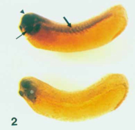
Fig. 2. Expression of CCTy in whole mount in situ preparations of
stage 30 Xenopus embryos. Hybridization with an antisense RNA probe
(top) identifies transcripts in the neural tube (arrowhead), pharyngeal
arches (thin arrow), and dorsolateral mesoderm (thick arrow). A sense
RNA probe (bottom) shows no specific cellular staining. There is faint
staining in the pharyngeal cavity c); histological analysis confirms that
this is not associated with cells (not shown). Image published in: Dunn MK and Mercola M (1996) Copyright © 1996. Image reproduced with permission of the Publisher, John Wiley & Sons.
Image source: Published Larger Image Printer Friendly View |
