XB-IMG-83364
Xenbase Image ID: 83364
|
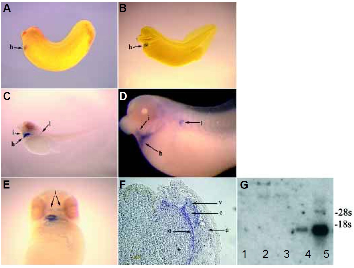
Fig. 2. Expression of MLP during Xenopus embryogenesis. (A) MLP expression is first detected at stage 27 (staged according to Nieuwkoop and Faber, 1994) in the heart (h) by whole mount in situ hybridization. (B) A stage 36 embryo showing that MLP expression (seen in blue) is still restricted to the heart at this stage. (C) At stage 40/41, MLP expression can be found in both the heart (h) and intermandibularis (i) muscle. In addition, expression was observed in the lymph heart (l) near the anterior somites. (D) An enlarged view of a stage 40/41 embryo showing the regions of expression in the head and clearly showing the lack of expression in the developing somites. (E) Ventral view of a Xenopus embryo showing the two patches of intermandibularis muscle spanning the lower jaw and expressing MLP. (F) A frontal section through the heart of a stage 38 embryo showing expression of MLP in the ventricle (v), atria (a) and outflow tracts (o) but not the endocardium (e). (G) Northern blot of adult intestine (1), kidney (2), liver (3), skeletal muscle (4), and heart (5) RNA probed with MLP. There was approximately equal loading in each lane based on rRNA levels. MLP expression is restricted to the heart and skeletal muscle. As in mammals, a higher level of expression was observed in the heart, in which a second, larger transcript was also observed (Arber et al., 1994). Image published in: Duan LJ et al. (2003) Copyright © 2003. Image reproduced with permission of the Publisher.
Image source: Published Larger Image Printer Friendly View |