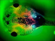
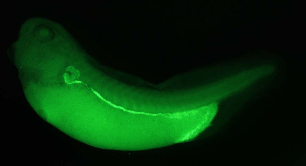
The pronephroi (plural of pronephros), also known as the head kidneys, are labeled blue in this sample due to the uptake of fluorescent proteins from the blood. Xenopus pronephroi leak large amounts of protein later recovered by transepithelial transport during glomeral filtration. See
Zhou and Vize (2004) for details. NF Stage 45, dorsal view, anterior left.
Pronephric tubule segment schematic and nomenclature. NF Stages 33 and 35. Copyright © 2004 Academic Press
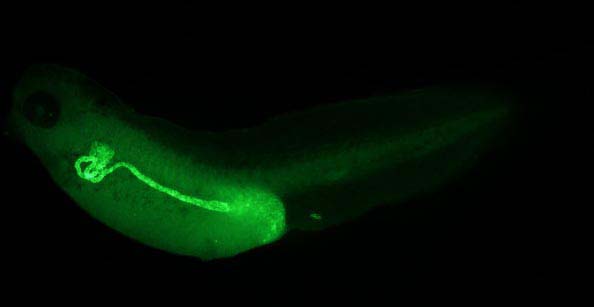
NF Stage 40: Sodium-potassium ATPase expression in pronephric tubules. Lateral view, anterior left.
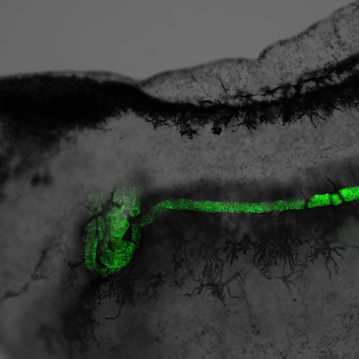
Sodium-potassium ATPase expression in pronephric tubules. Confocal micrograph combining fluorescent and transmitted light. Lateral view closeup.
Pronephric tubules labeled by FCIS with a sodium bicarnonate cotransporter and NaK ATPase. Copyright © 2004 Academic Press
Pronephric tubules labeled by FCIS with an amino acid transporter,
slc4a4, an NaK ATPase. Blue stain is in late proximal segment (also see section below). Copyright © 2004 Academic Press
Early distal segment labeled red and late distal segment labeled green.
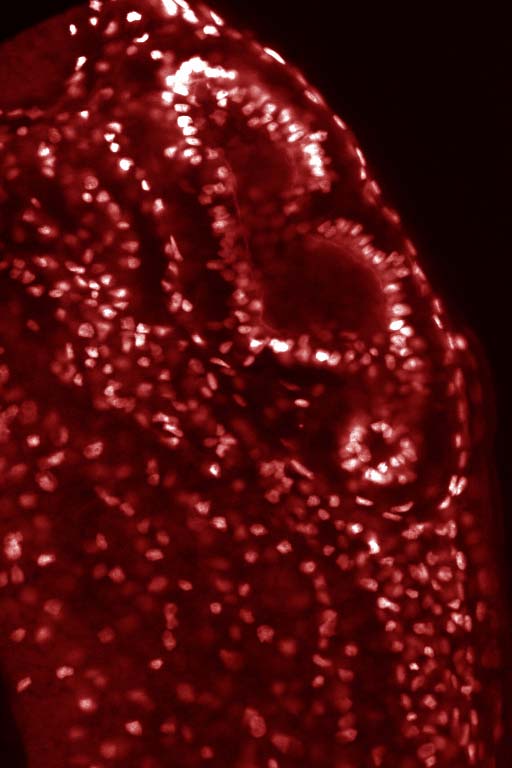
Stage 35 pronephric tubules: lateral view, anterior to the left. Propidium iodide staining.
Wallingford et al. 1998. Copyright © 1998 Academic Press.
Stage 35 pronephros, transverse section. Propidium iodide staining.
Wallingford et al. 1998 . Copyright © 1998 Academic Press.
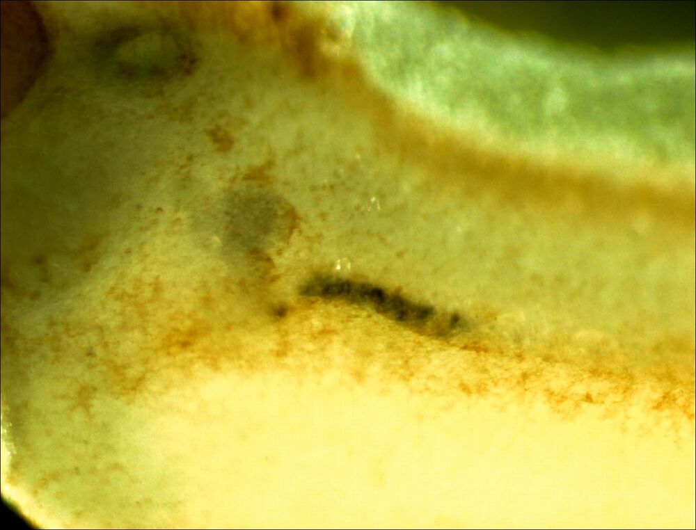
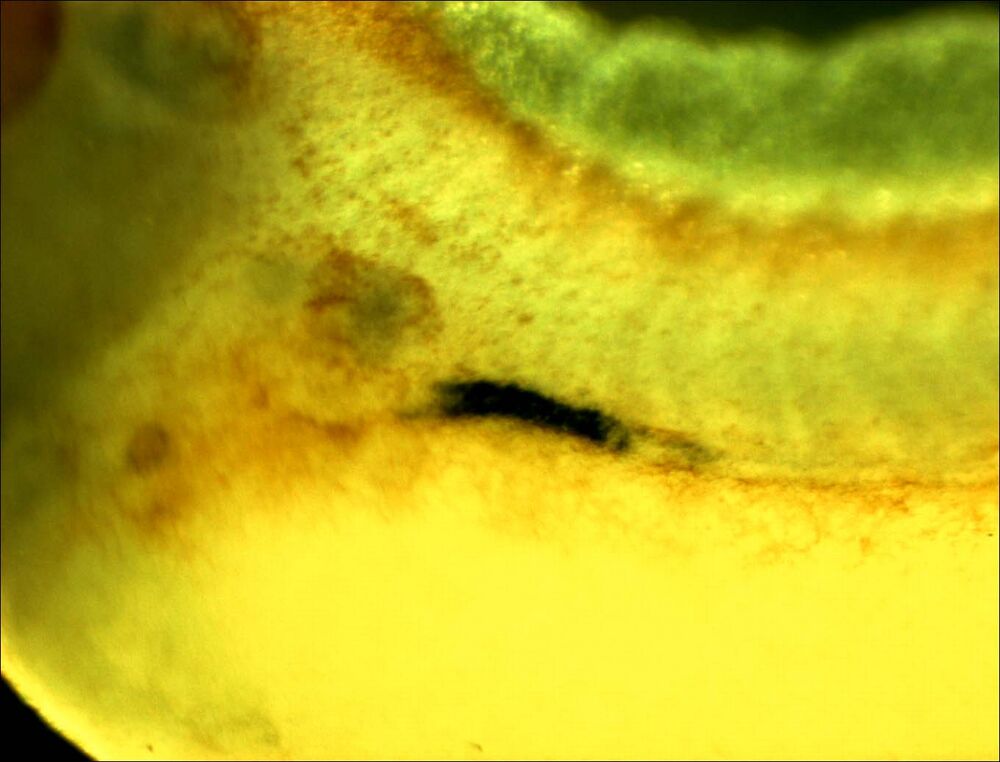
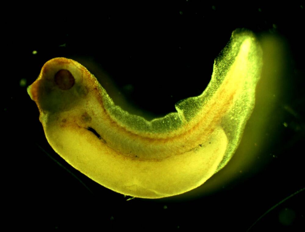
Additional images highlighting slc4a4:
NF Stage 31, lateral view. Early phase of expression restricted to the proximal segment.
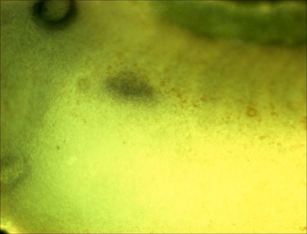
NF Stage 34 embryo, close up. Both light proximal staining and dark late distal staining visible.
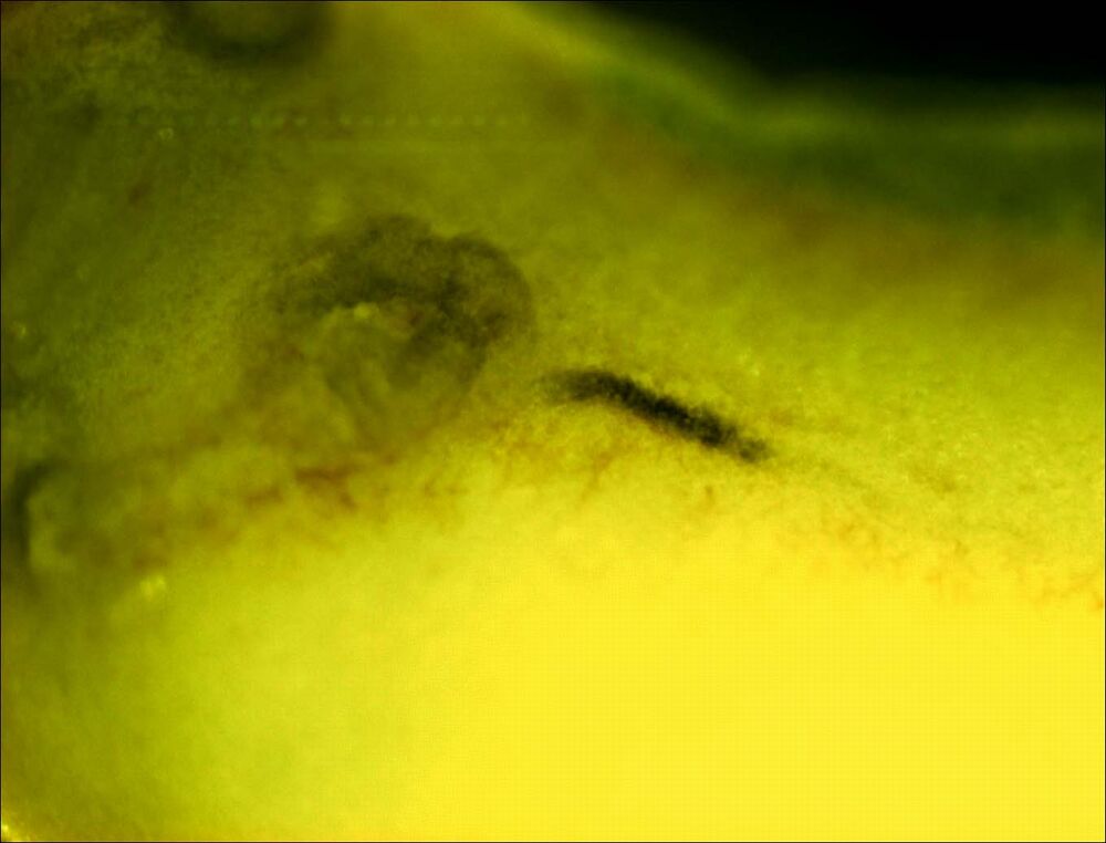
NF Stage 35 embryo: slc4a4 staining.
NF Stage 38 embryo: slc4a4 staining.
NF Stage 38-39 embryo: slc4a4 staining, full lateral view, anterior left.
NF Stage 35 staining. slc4a4 is shown in black, the NAK ATPase in FITC (green).
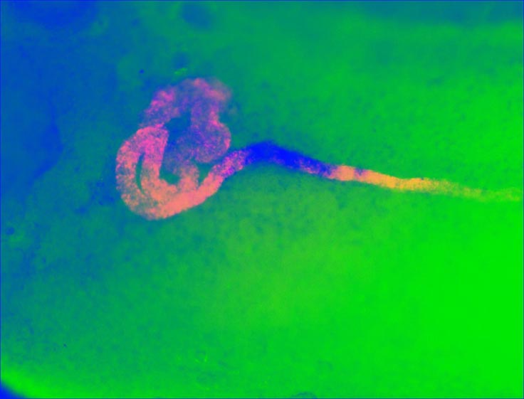
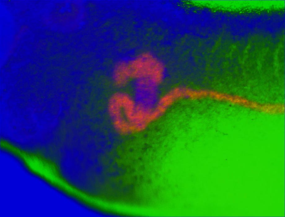
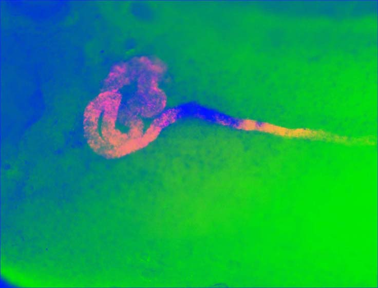
NF Stage 36 double stain. FCIS overlay. slc4a4 appears blue. ATPase alone appears orange. Overlap is pink.
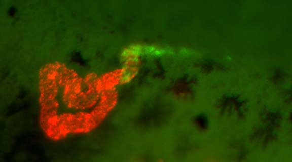
NF Stage 38 double stain. slc4a4 is shown in green (FITC), and XNKCC2 in red (Cy3). Wholemount FISH.
Adapted with permission from Disease Models & Mechanisms: Blackburn & Miller. (2019). Modeling congenital kidney diseases in Xenopus laevis. Dis Model Mech. 2019 Apr 9;12(4). pii: dmm038604. doi: 10.1242/dmm.038604. Copyright (2019).
This work is licensed under a Creative Commons Attribution 4.0 International License. The images or other third party material in this article are included in the article’s Creative Commons license, unless indicated otherwise in the credit line; if the material is not included under the Creative Commons license, users will need to obtain permission from the license holder to reproduce the material. To view a copy of this license, visit http://creativecommons.org/licenses/by/4.0/


