Early development: X. laevis histology
The Early Development of Xenopus laevis: An Atlas of the Histology
Peter Hausen and Metta Riebsell
1991
Verlag der Zeitschrift für Naturforschung
ISBN: 0-387-53740-6
Images reproduced on Xenbase with permission from the publisher. Contact publisher for permission to reproduce images.
Images copyright Verlag der Zeitschrift für Naturforschung (1991).
Publisher URL.
[click image to view enlargement]
The Early Development
of Xenopus laevis
Hausen and Riebsell
Book cover.
of Xenopus laevis
Hausen and Riebsell
Book cover.
Oogenesis
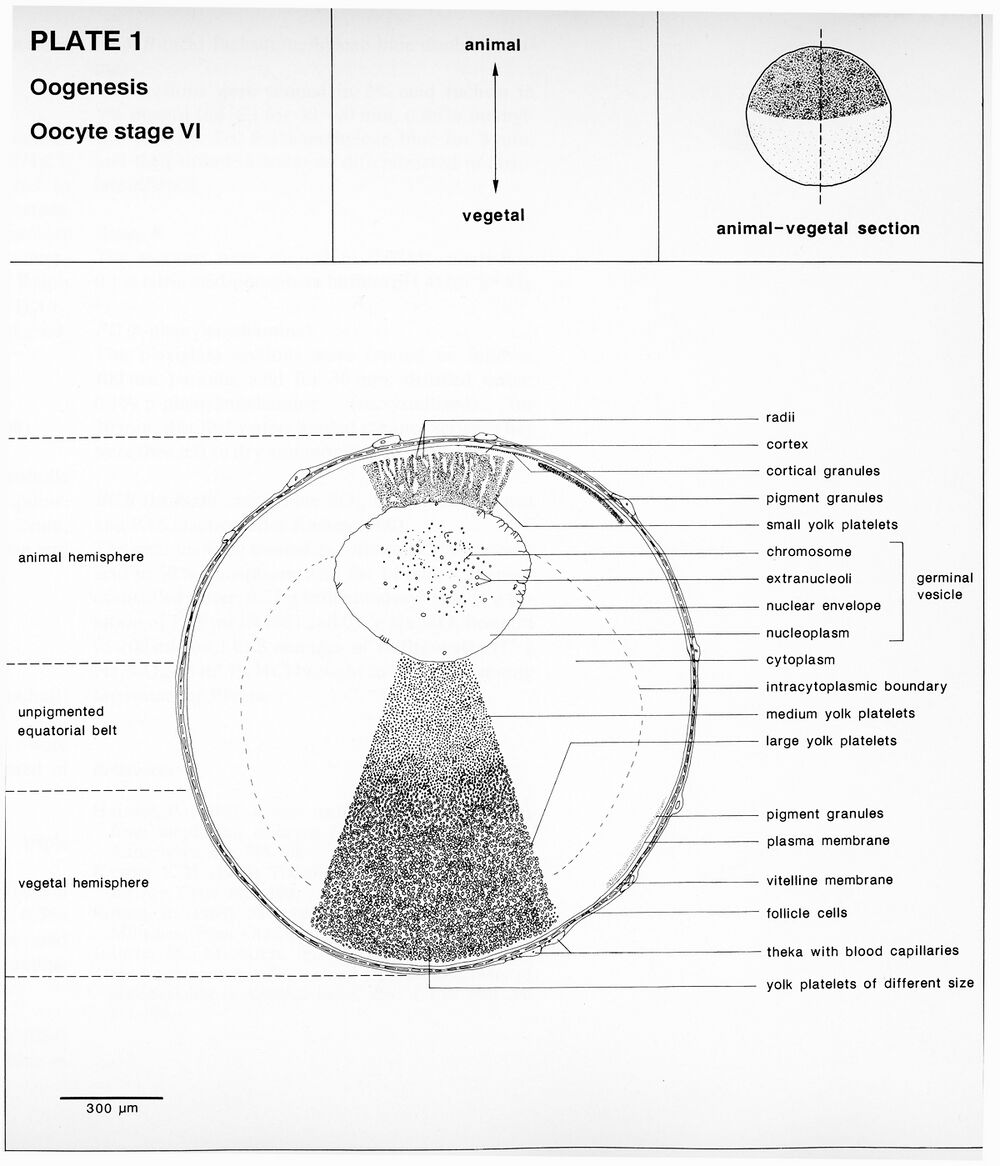
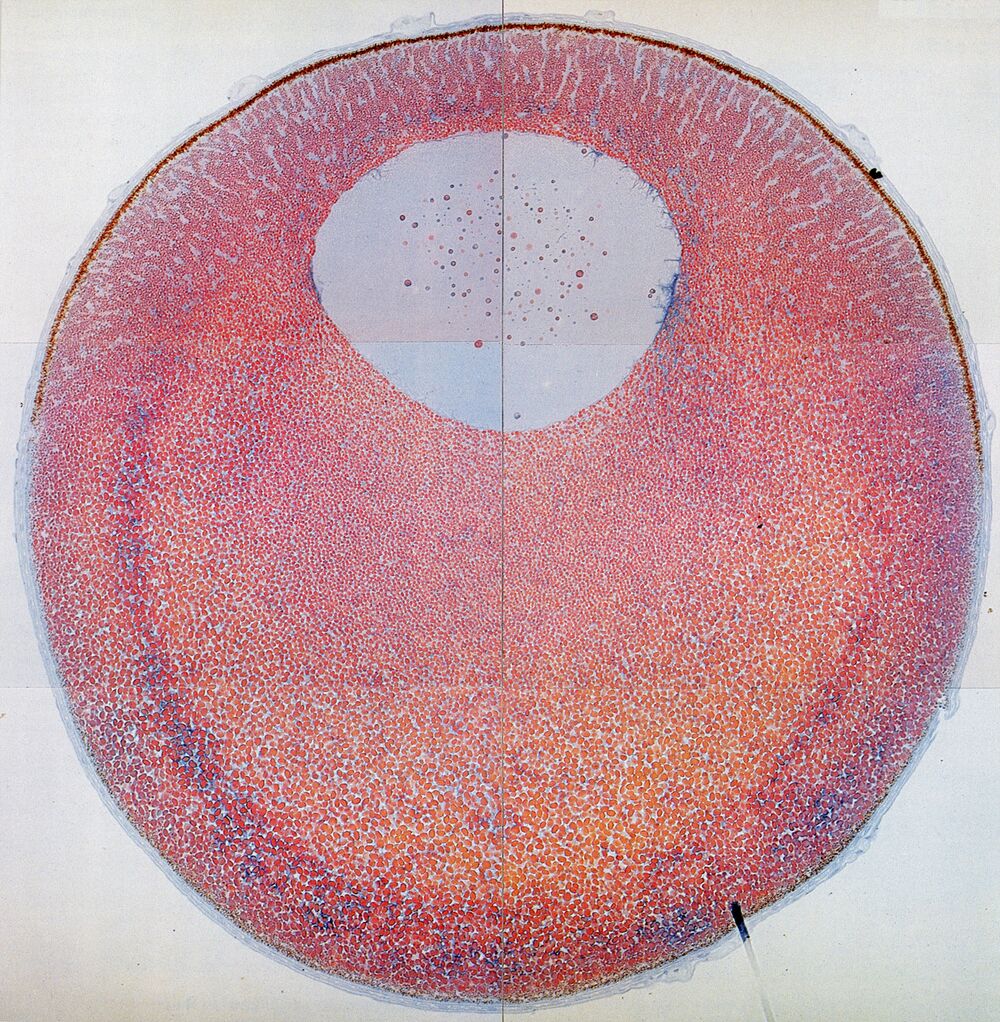
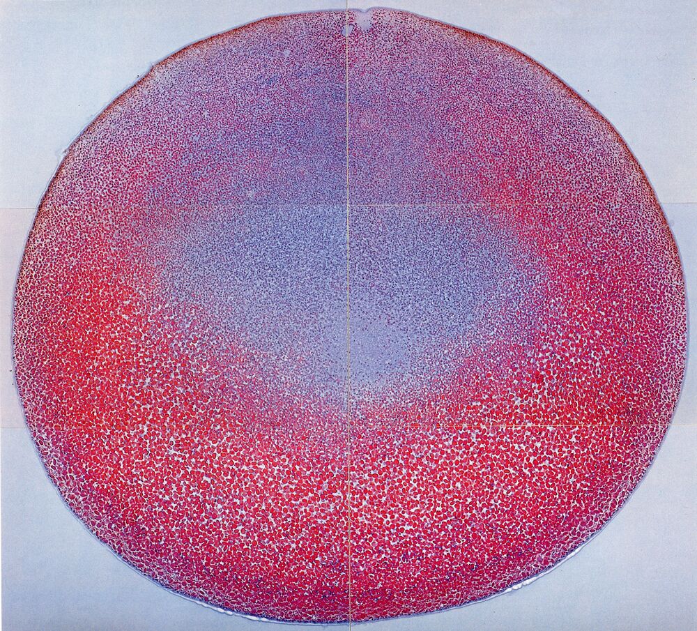
Plate 1: Oogenesis, oocyte stage VI. The fully grown oocyte.
Higher resolution Plate 1.
Higher resolution Plate 1.
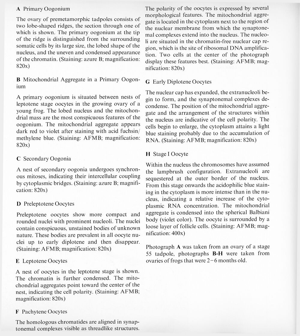
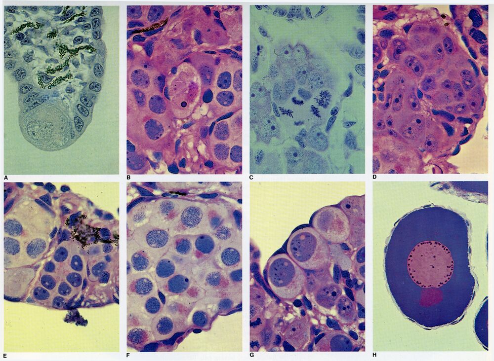
Plate 2: Oogenesis: Premeiotic oogonia and oocyte in early meiotic prophase.
Higher resolution Plate 2.
Sections 2a, 2b, 2c, 2d, 2e, 2f, 2g, and 2h.
Higher resolution Plate 2.
Sections 2a, 2b, 2c, 2d, 2e, 2f, 2g, and 2h.
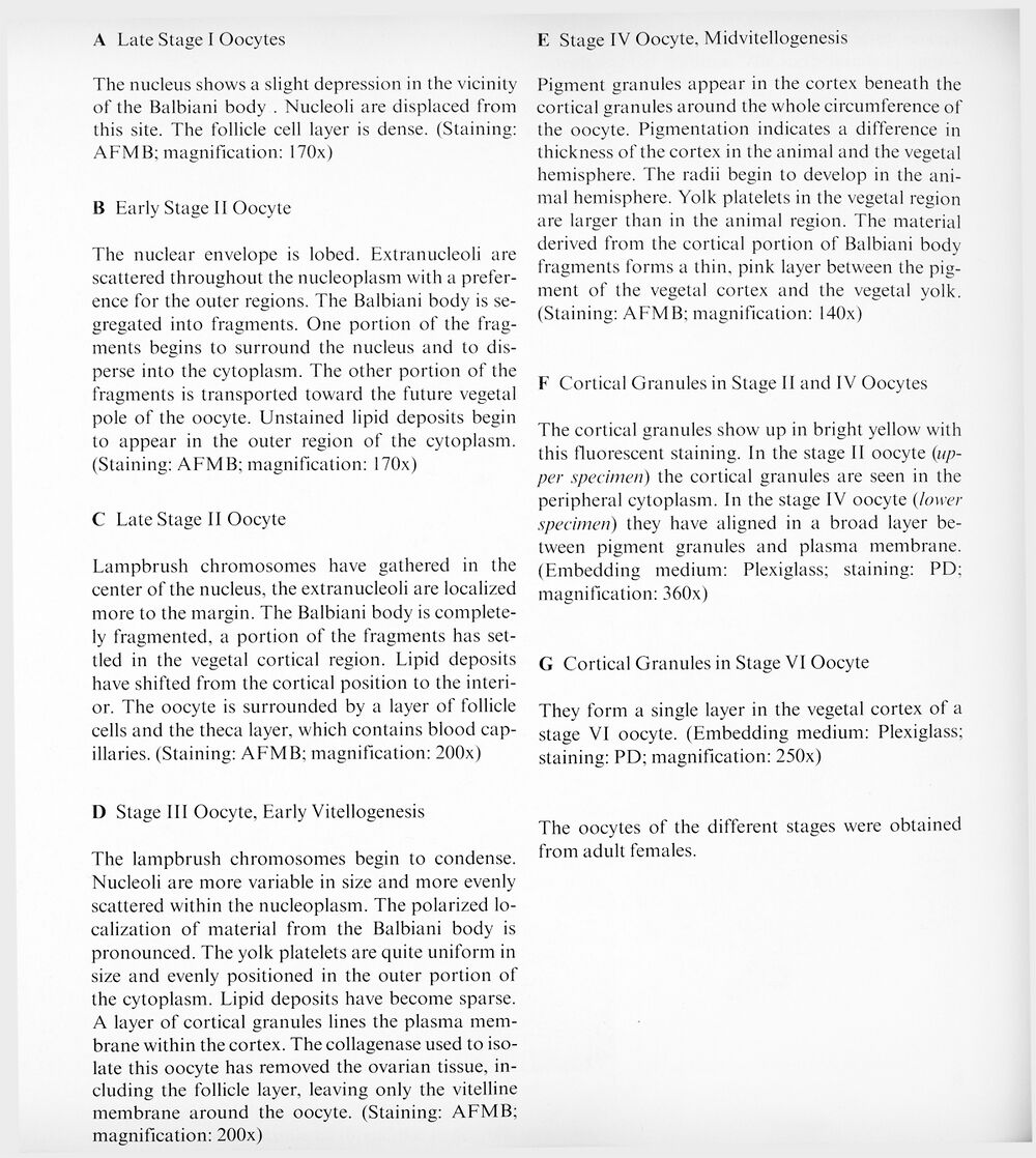
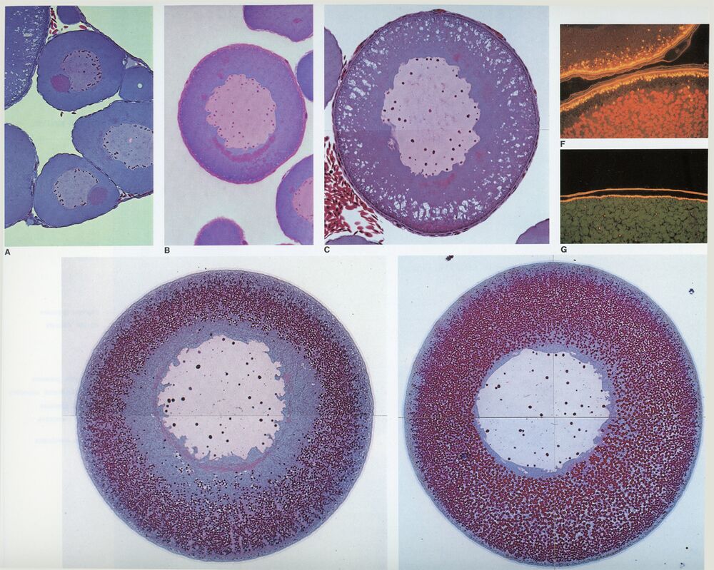
Plate 3: Oogenesis: Oocytes, Stages I-IV.
Higher resolution Plate 3.
Sections 3a, 3b, 3c, 3d, 3e, 3f, and 3g.
Higher resolution Plate 3.
Sections 3a, 3b, 3c, 3d, 3e, 3f, and 3g.
Oocyte maturation
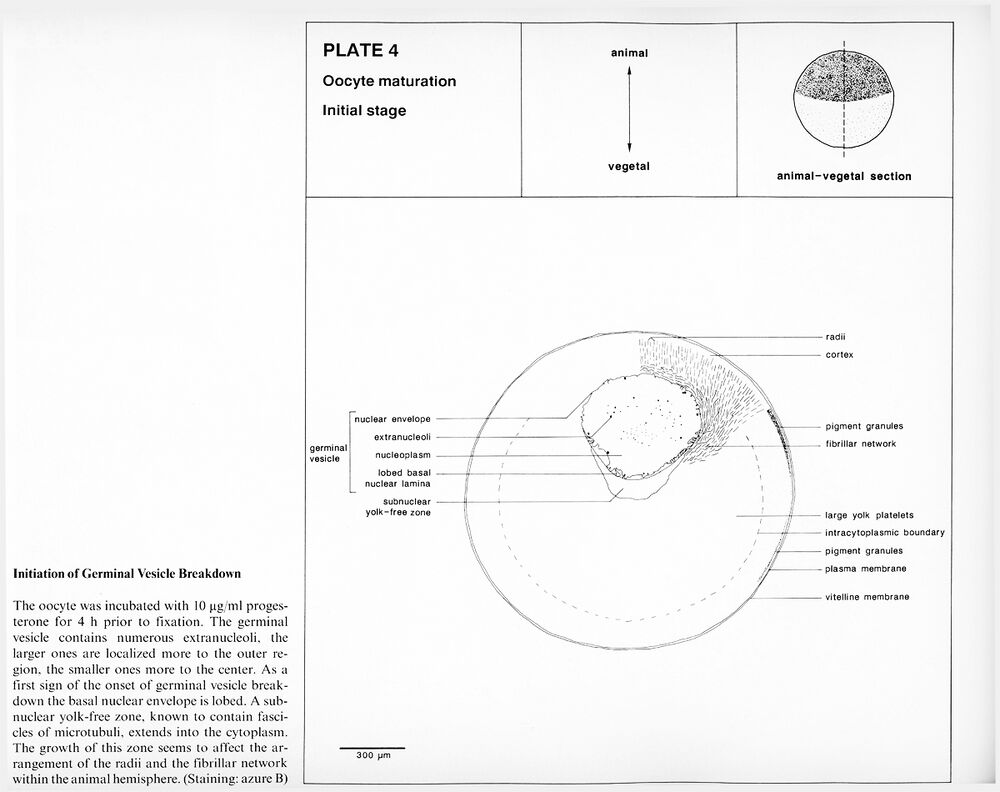
Plate 4: Initiation of germinal vesicle breakdown.
Higher resolution Plate 4.
Higher resolution Plate 4.
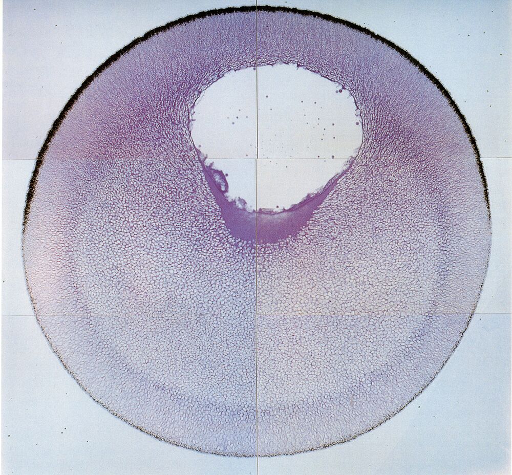
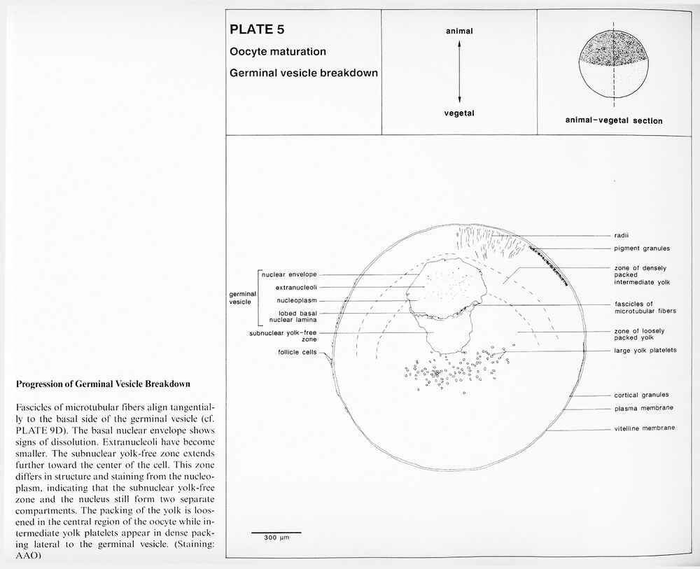
Plate 5: Progression of germinal vesicle breakdown.
Higher resolution Plate 5.
Higher resolution Plate 5.
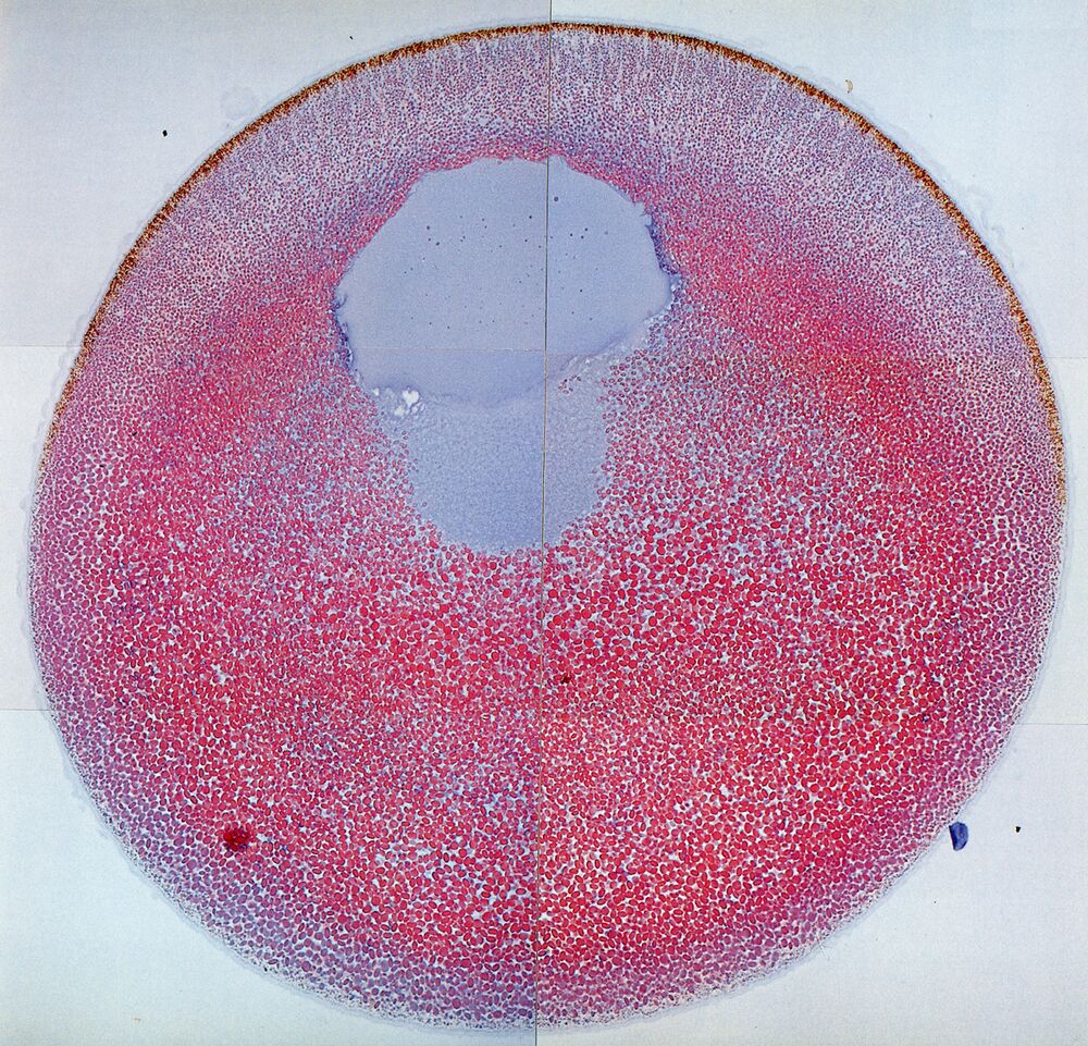
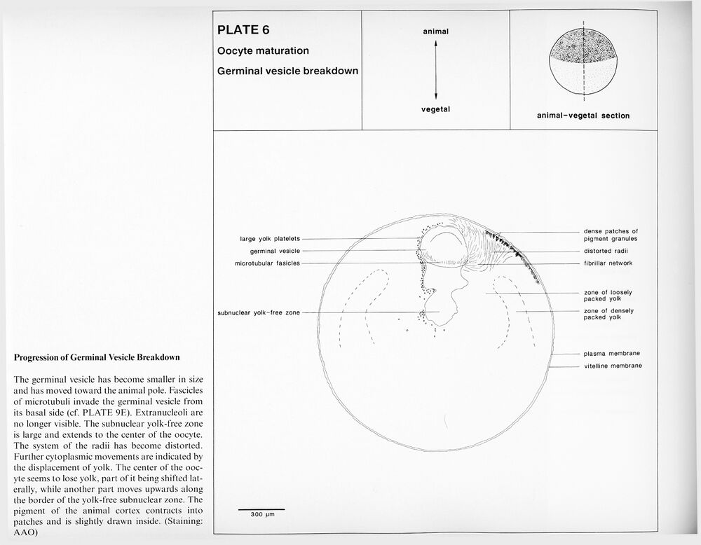
Plate 6: Progression of germinal vesicle breakdown.
Higher resolution Plate 6.
Higher resolution Plate 6.
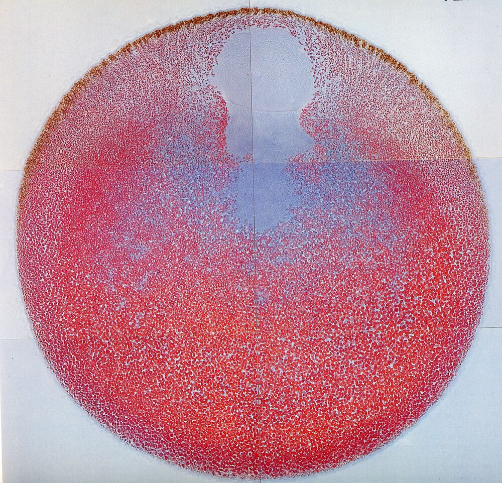
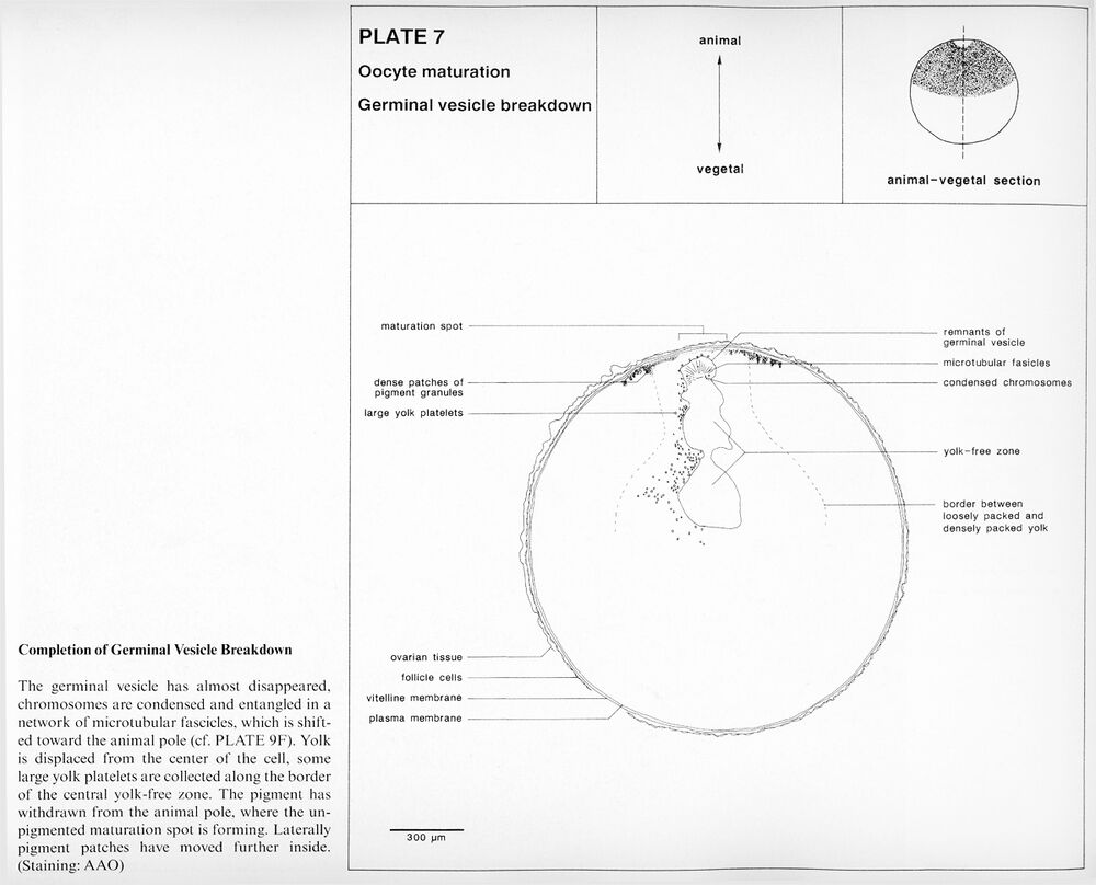
Plate 7: Completion of germinal vesicle breakdown.
Higher resolution Plate 7.
Higher resolution Plate 7.
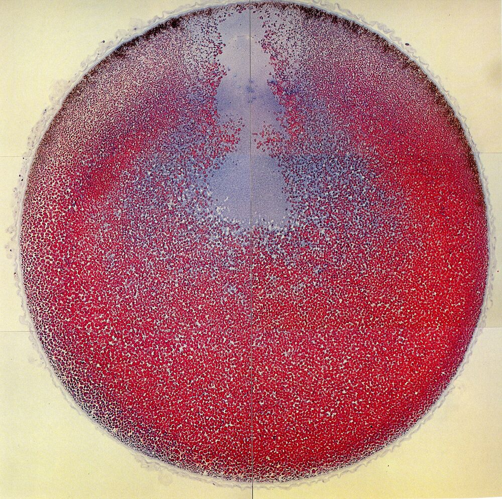
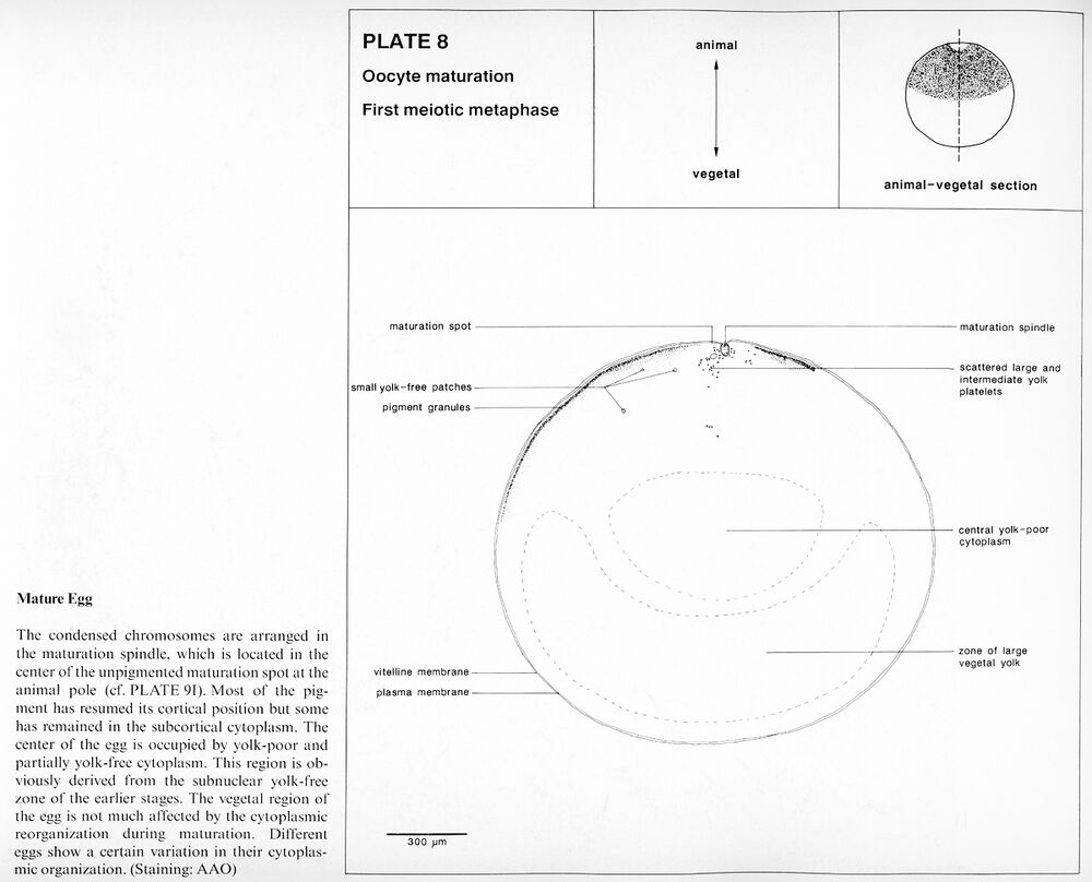
Plate 8: Mature egg, first meotic metaphase.
Higher resolution Plate 8.
Higher resolution Plate 8.
Oogenesis and Maturation
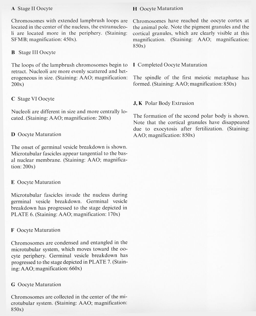
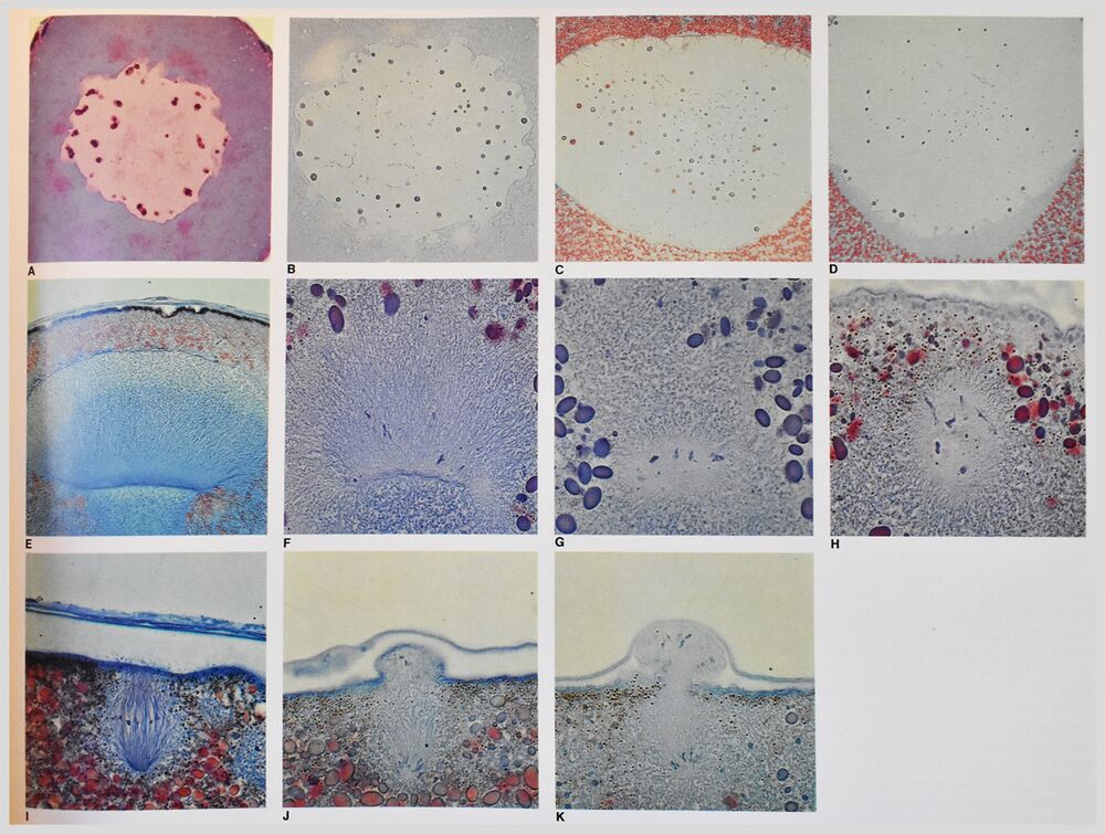
Plate 9: Nuclear events during meiosis at higher magnification
Higher resolution Plate 9.
Sections 9a, 9b, 9c, 9d, 9e, 9f, 9g, 9h, 9i, 9j, and 9k.
Higher resolution Plate 9.
Sections 9a, 9b, 9c, 9d, 9e, 9f, 9g, 9h, 9i, 9j, and 9k.
Fertilized egg
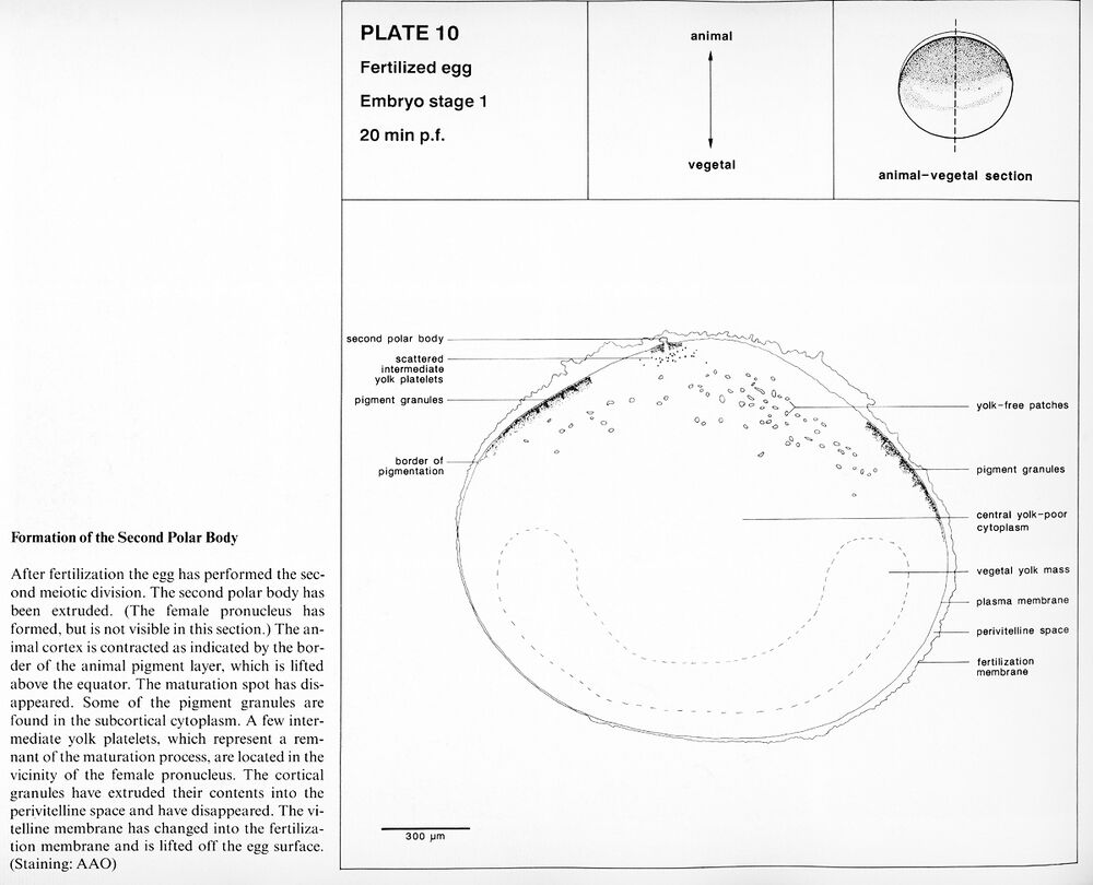
Plate 10: Embryo stage 1, Formation of the second polar body. 20 min. p.f.
Higher resolution Plate 10.
Higher resolution Plate 10.
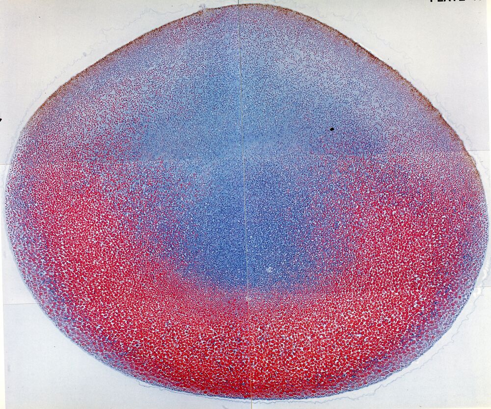
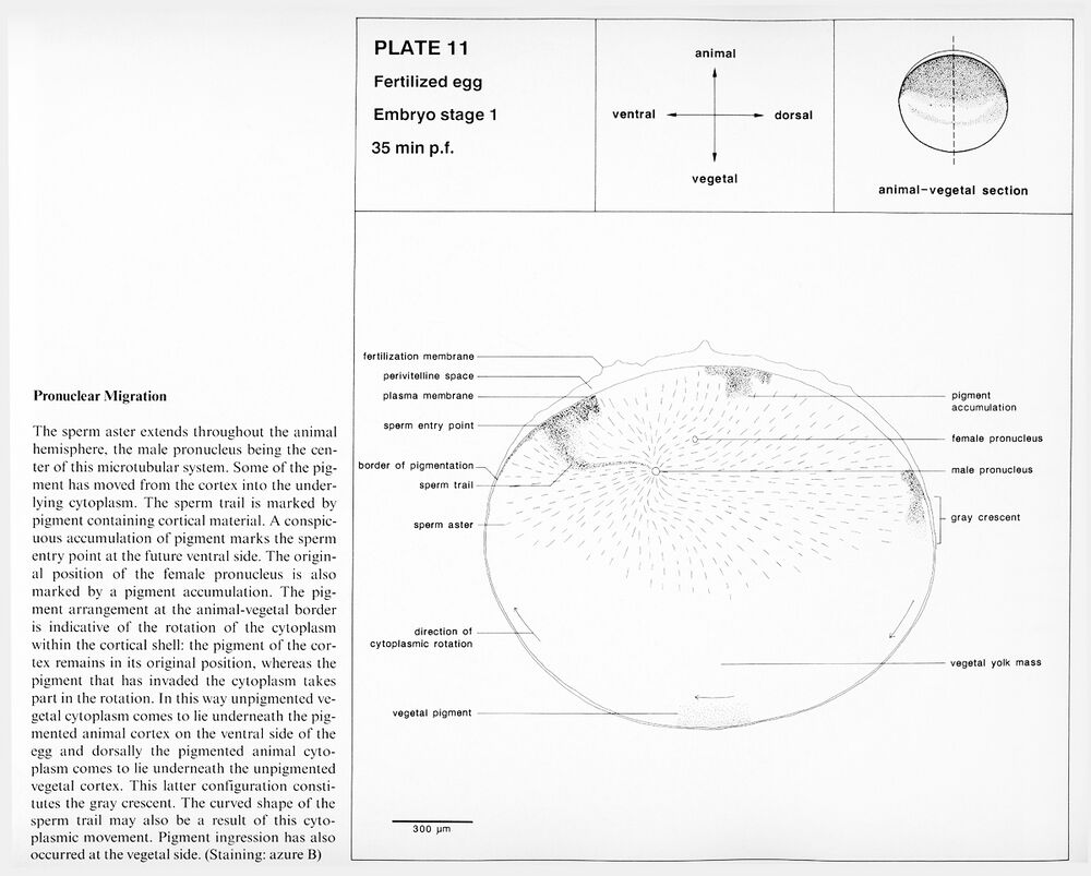
Plate 11: Embryo stage 1, Pronuclear migration. 35 min. p.f.
Higher resolution Plate 11.
Higher resolution Plate 11.
Plate 12: Embryo stage 1, Karyogamy. 45 min. p.f.
Higher resolution Plate 12.
Higher resolution Plate 12.
First cleavage
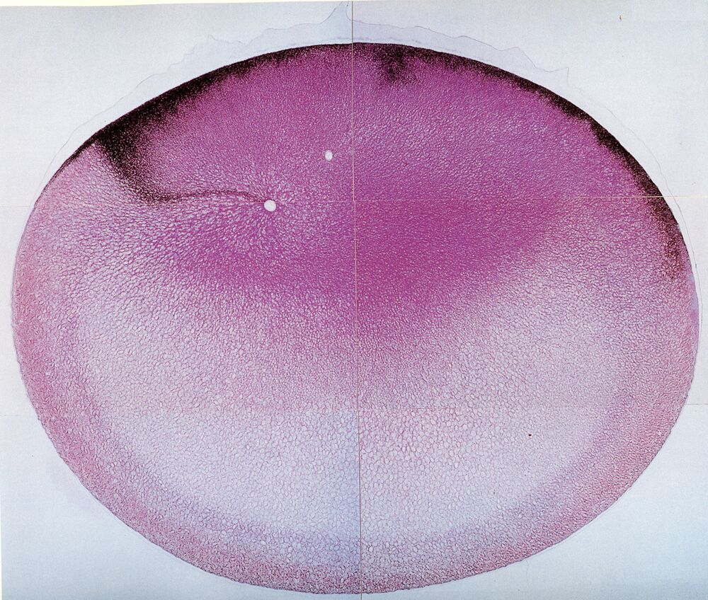
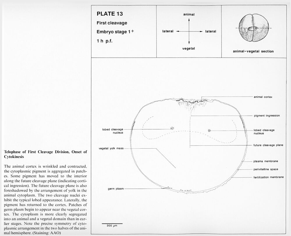
Plate 13: Embryo stage 1+, Telophase of first cleavage division, onset of cytokinesis. 1 h p.f.
Higher resolution Plate 13.
Higher resolution Plate 13.
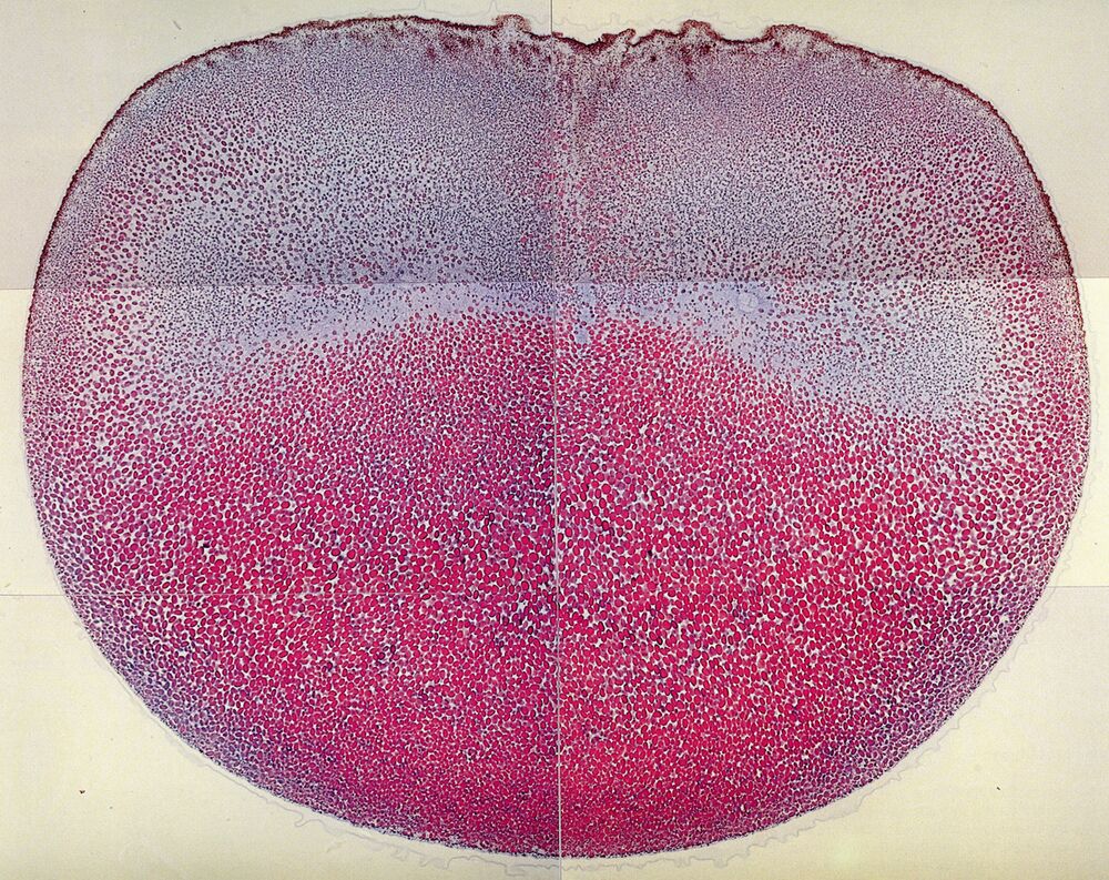
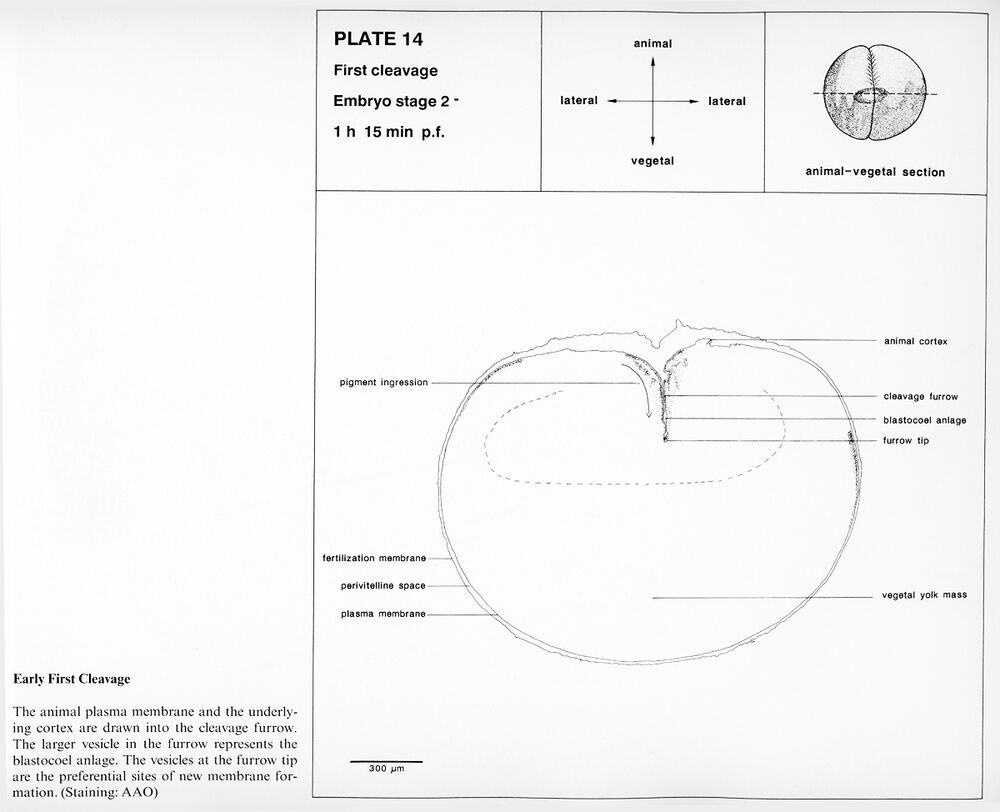
Plate 14: Embryo stage 2-, Early first cleavage. 1 h 15 min p.f.
Higher resolution Plate 14.
Higher resolution Plate 14.
2-cell stage
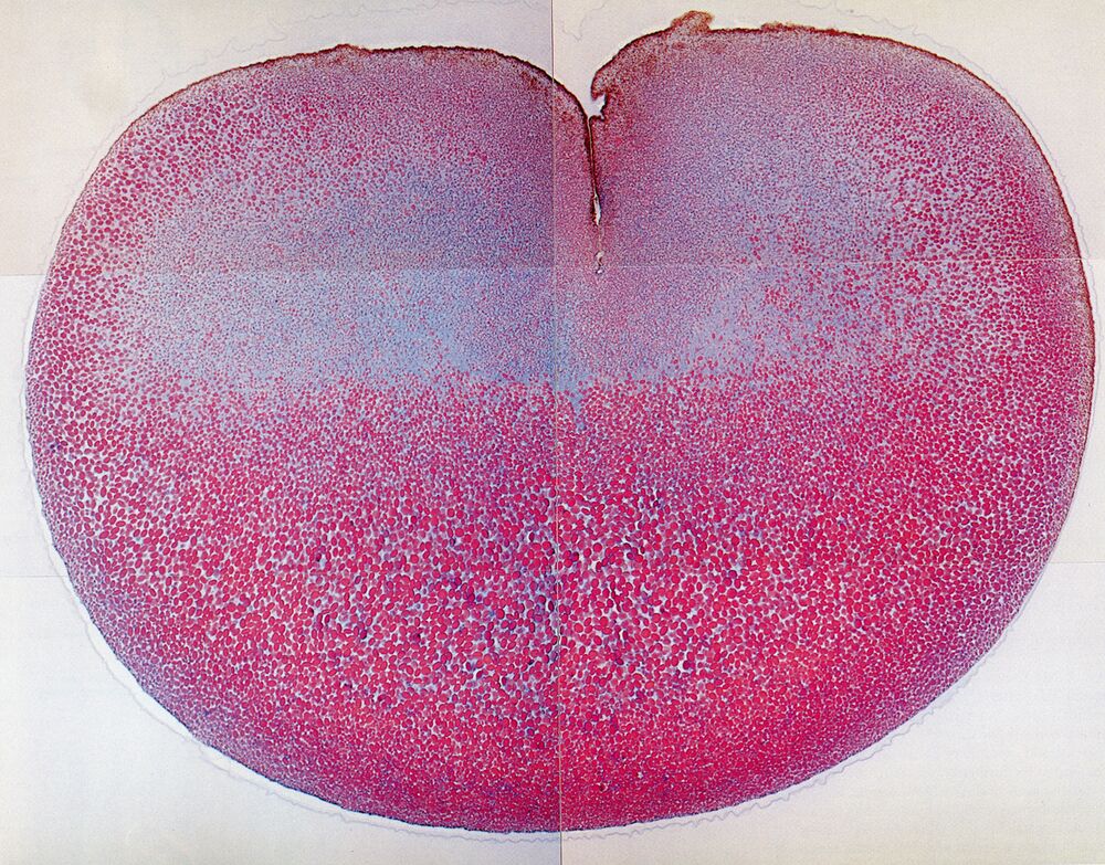
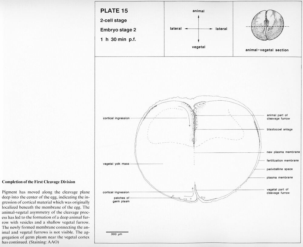
Plate 15: Embryo stage 2, Completion of first cleavage division. 1 h 30 min p.f.
Higher resolution Plate 15.
Higher resolution Plate 15.
8-cell stage
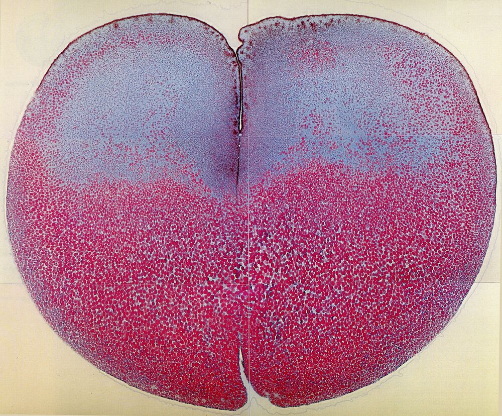
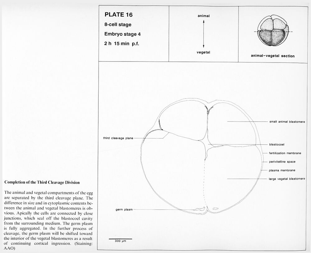
Plate 16: Embryo stage 4, Completion of the third cleavage division. 2 h 15 min p.f.
Higher resolution Plate 16.
Higher resolution Plate 16.
Fine-cell blastula
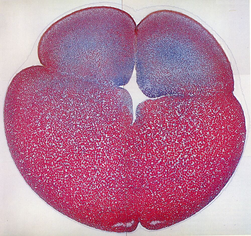
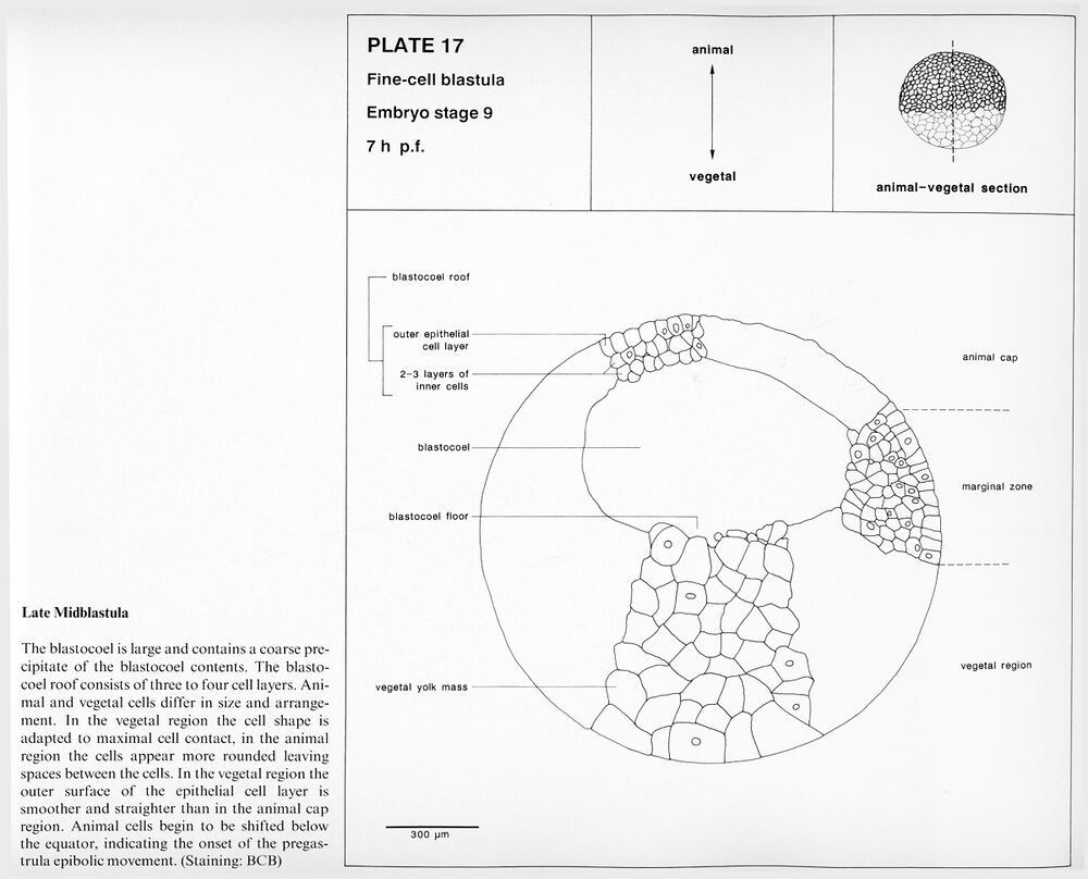
Plate 17: Embryo stage 9, Late midblastula. 7 h p.f.
Higher resolution Plate 17.
Higher resolution Plate 17.
Early gastrula
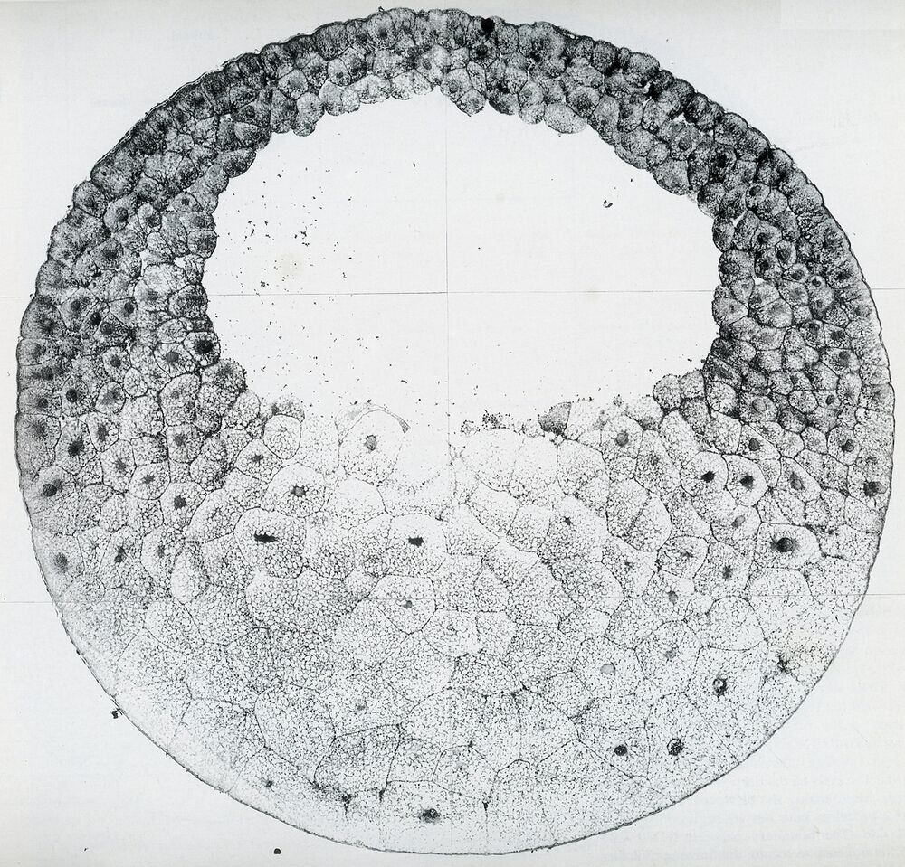
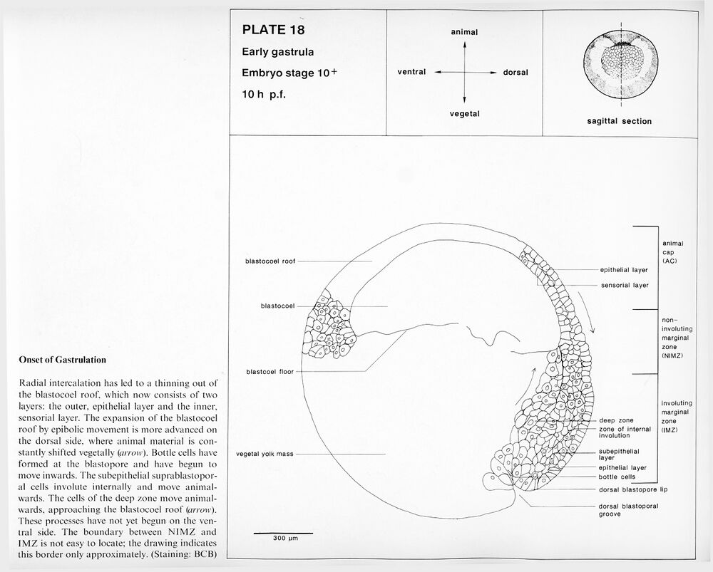
Plate 18: Embryo stage 10+, Onset of gastrulation. 10 h p.f.
Higher resolution Plate 18.
Higher resolution Plate 18.
Midgastrula
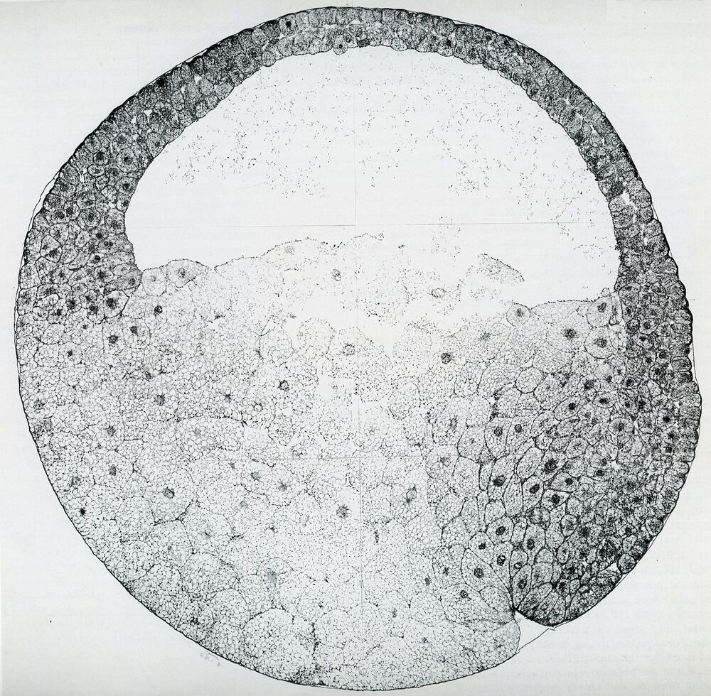
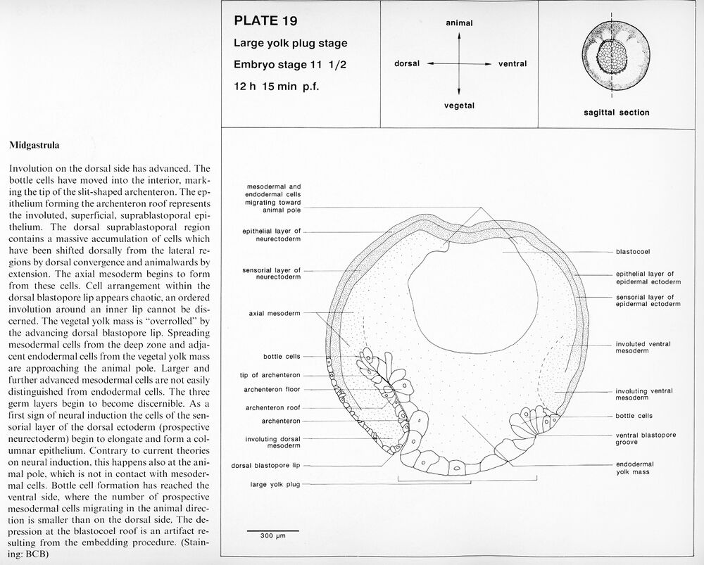
Plate 19: Embryo stage 11.5, Large yolk plug stage. 12 h 15 min p.f.
Higher resolution Plate 19.
Higher resolution Plate 19.
Advanced gastrula
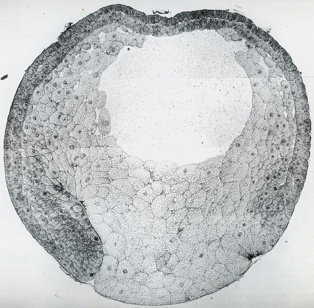
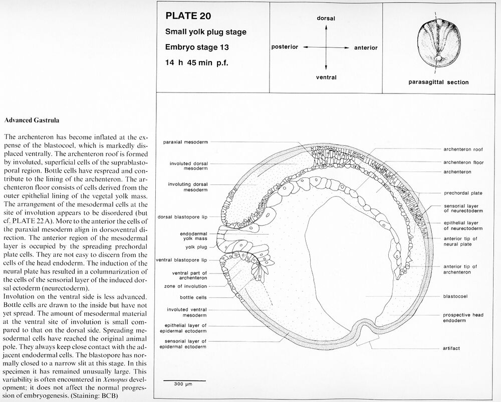
Plate 20: Embryo stage 13, Small yolk plug stage. 14 h 55 min p.f.
Higher resolution Plate 20.
Higher resolution Plate 20.
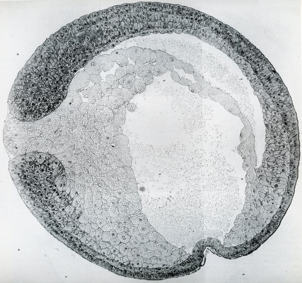
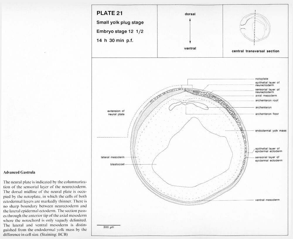
Plate 21: Embryo stage 12.5, Small yolk plug stage. 14 h 30 min p.f.
Higher resolution Plate 21.
Higher resolution Plate 21.
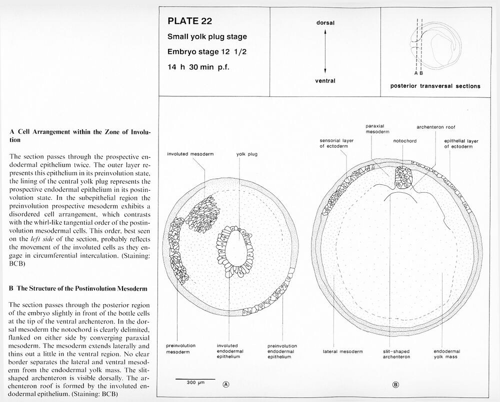
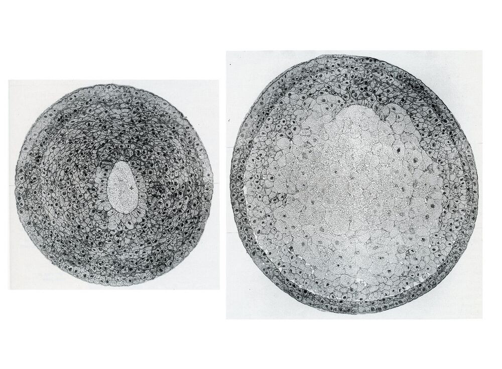
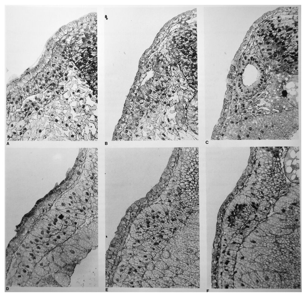
Plate 22: Embryo stage 12.5, Small yolk plug stage. 14 h 30 min p.f. A. Cell arrangement within the zone of involution. B. The structure of the postinvolution mesoderm.
Higher resolution plate sections 22a and 21b.
Higher resolution plate sections 22a and 21b.
Neural plate stage
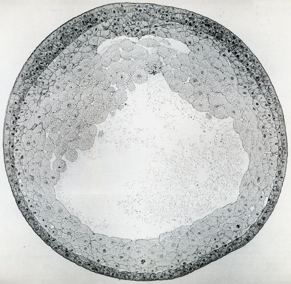
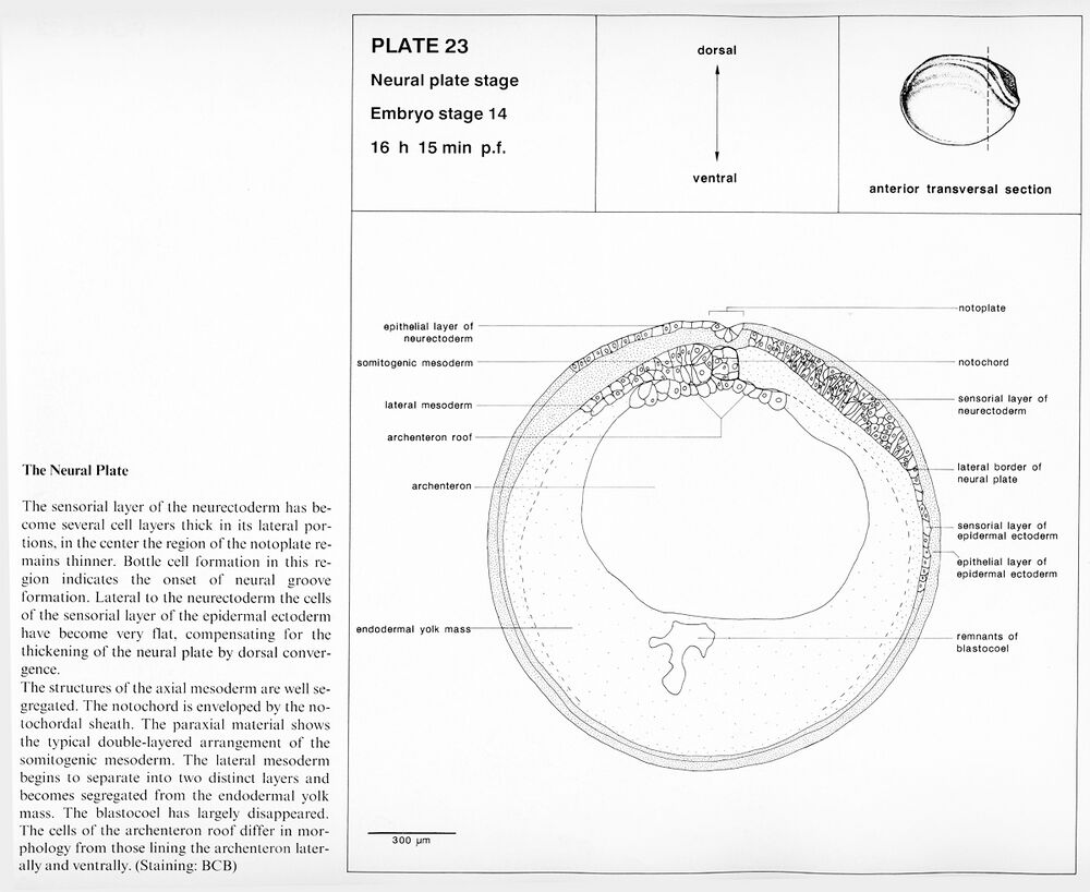
Plate 23: Embryo stage 14, The neural plate. 16 h 15 min p.f.
Higher resolution Plate 23.
Higher resolution Plate 23.
Neurula
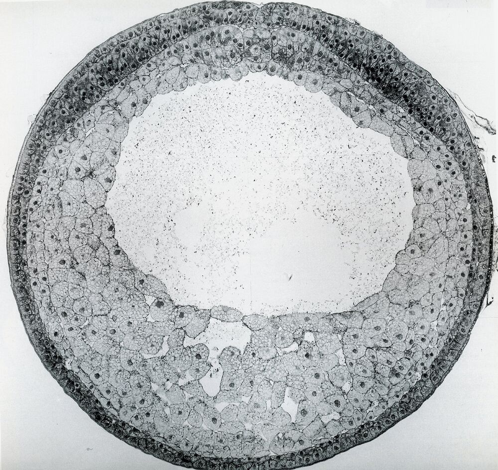
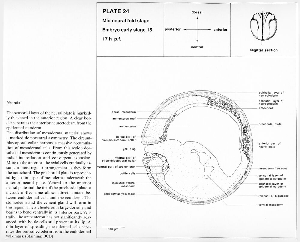
Plate 24: Embryo stage 15, Mid neural fold stage. 17 h p.f.
Higher resolution Plate 24.
Higher resolution Plate 24.
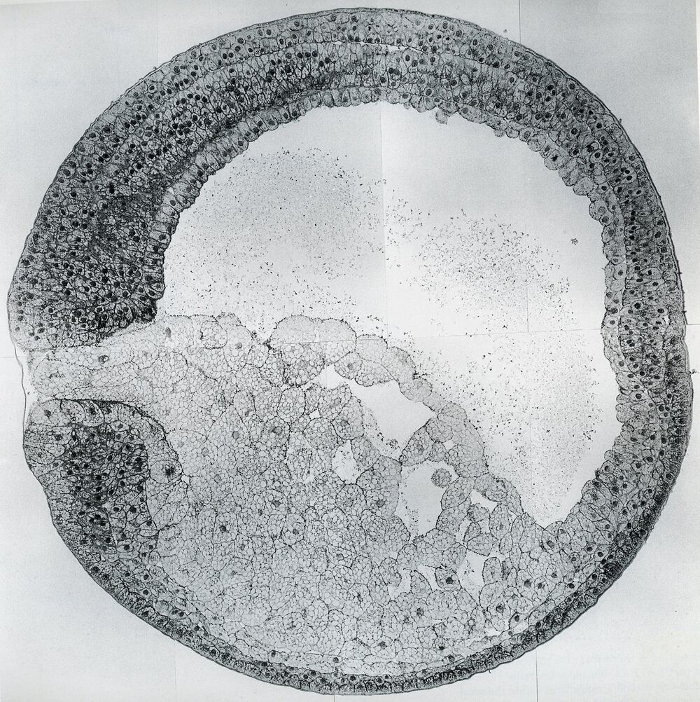
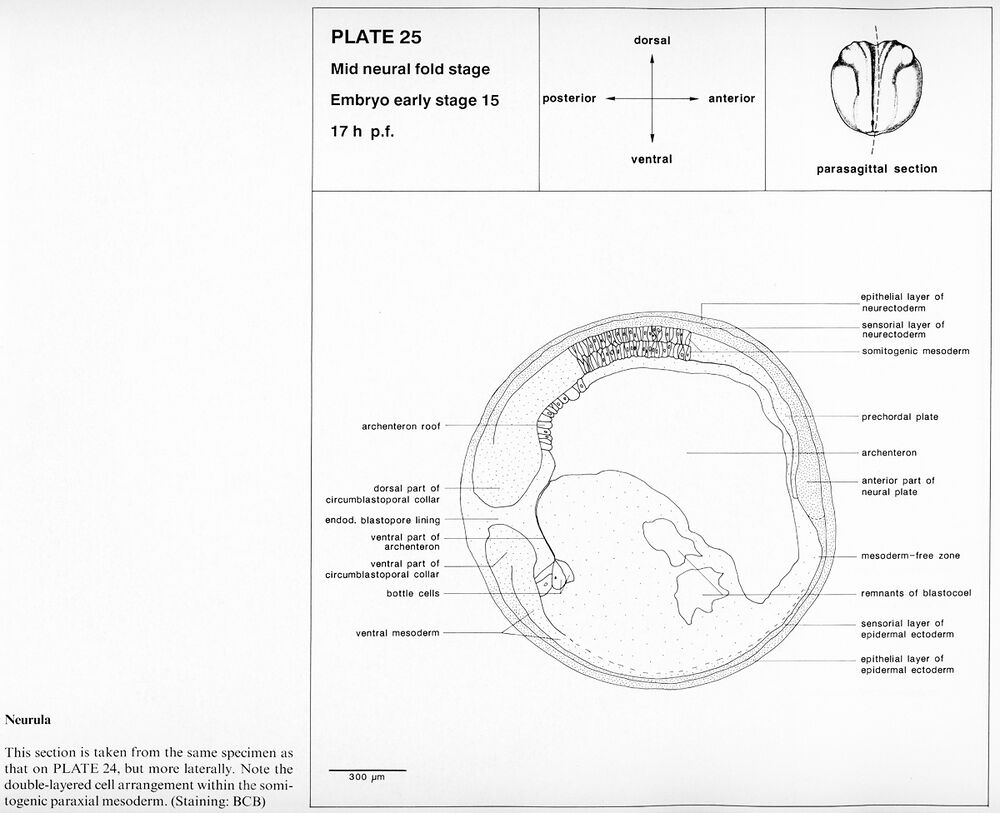
Plate 25: Embryo stage 15, Mid neural fold stage. 17 h p.f.
Higher resolution Plate 25.
Higher resolution Plate 25.
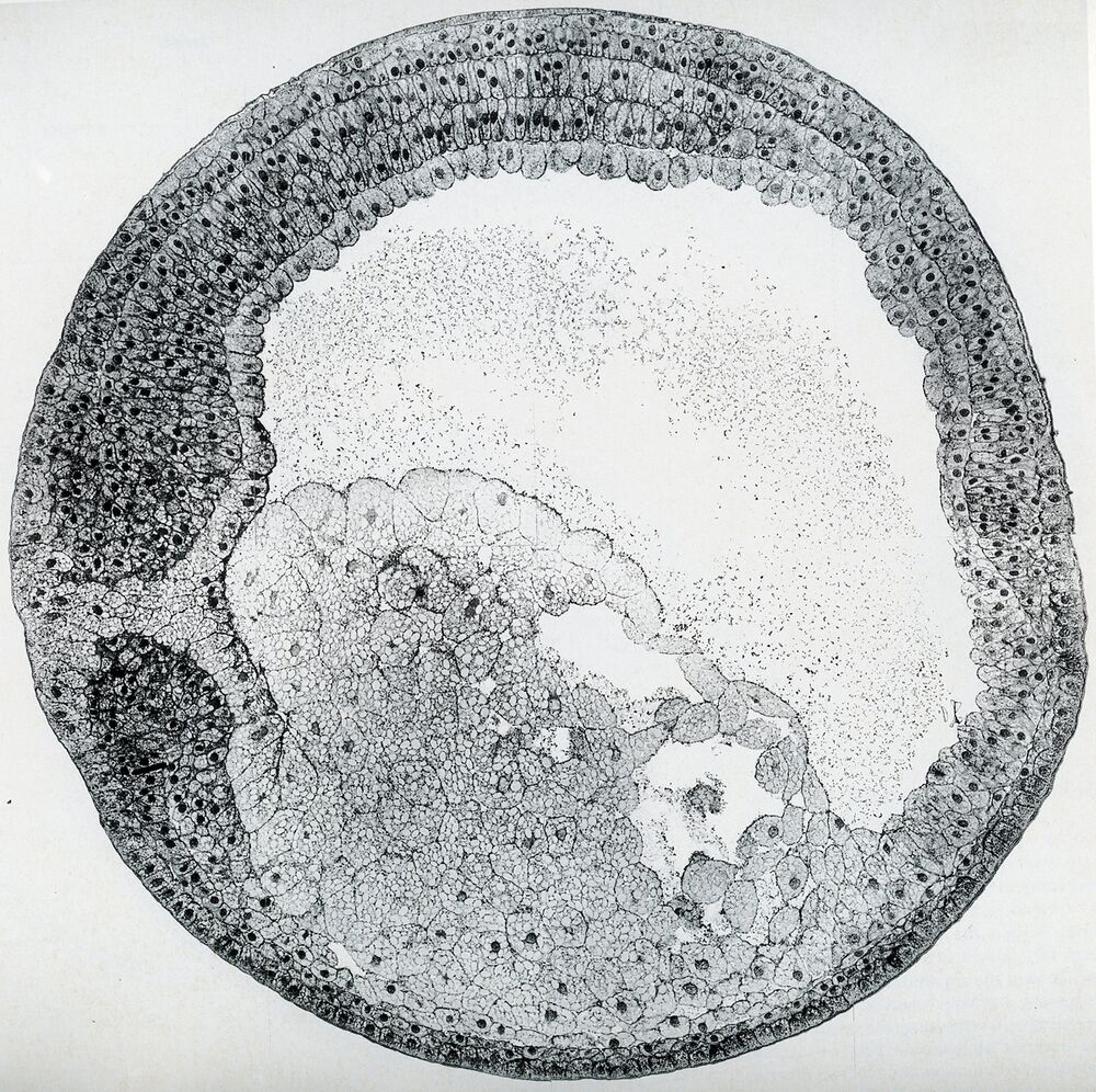
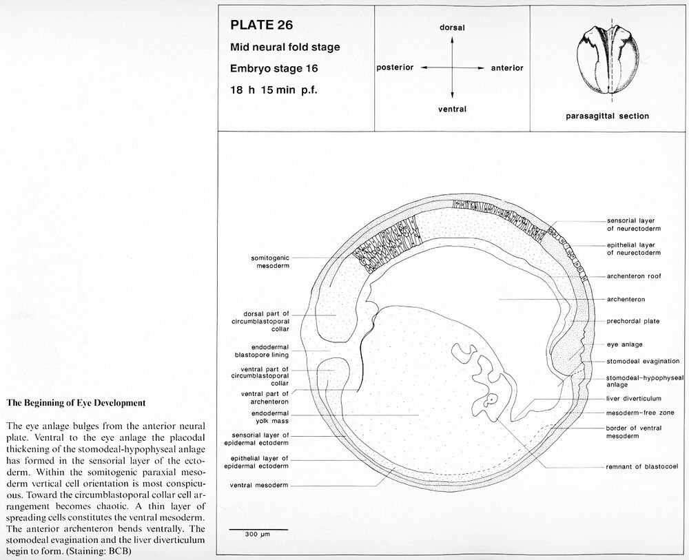
Plate 26: Embryo stage 16, Mid neural fold stage. The beginning of eye development. 18 h 15 min p.f.
Higher resolution Plate 26.
Higher resolution Plate 26.
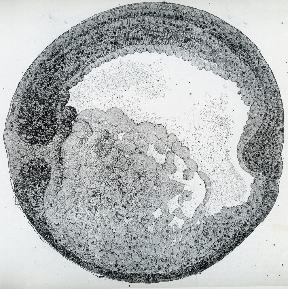
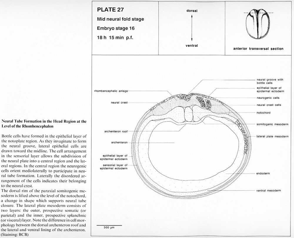
Plate 27: Embryo stage 16, Mid neural fold stage. Neural tube formation in the head region at the level of the rhombencephalon. 18 h 15 min p.f.
Higher resolution Plate 27.
Higher resolution Plate 27.
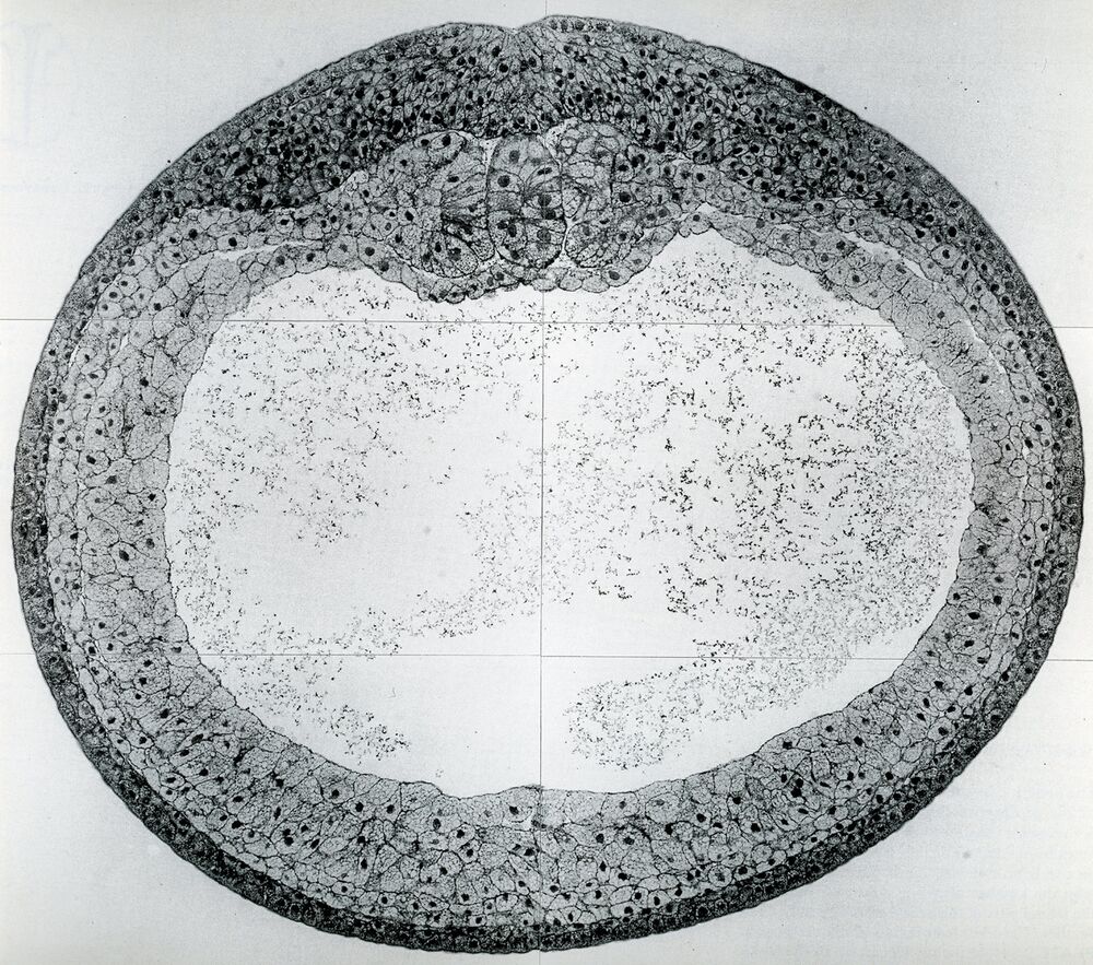
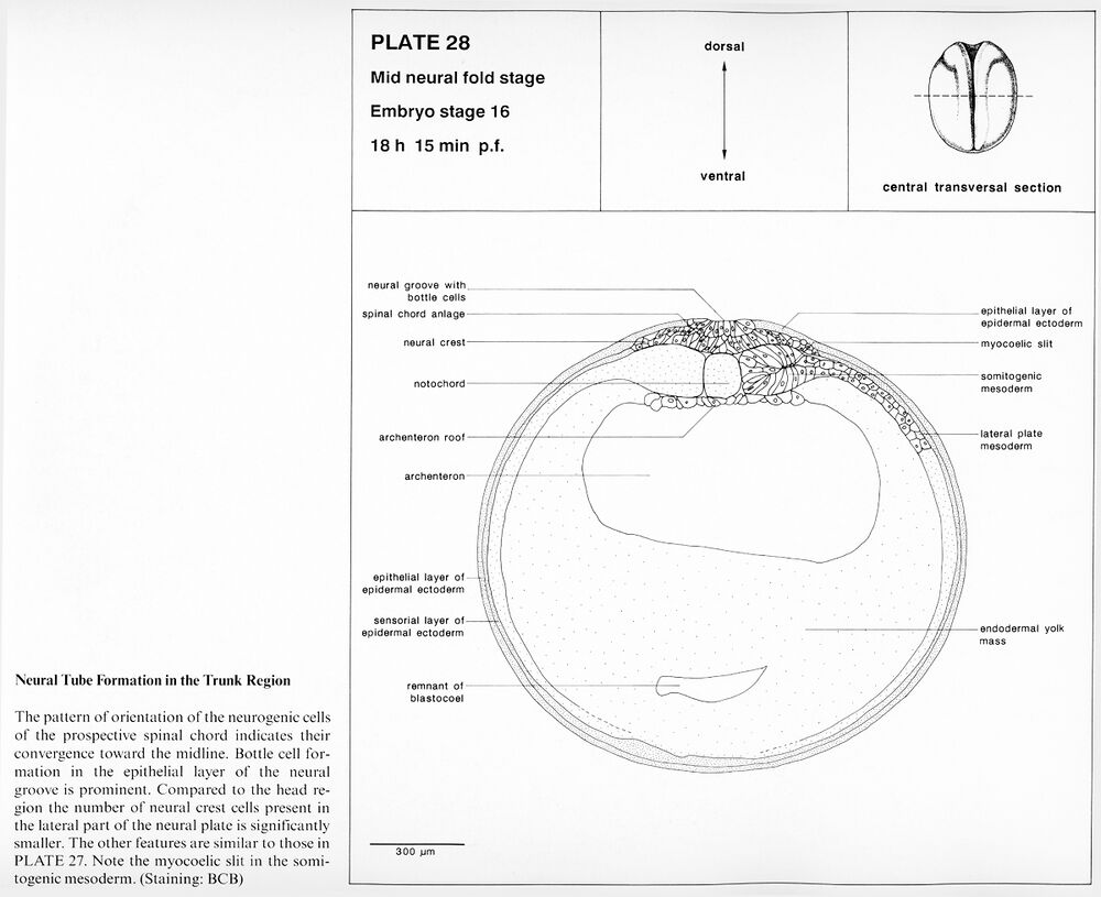
Plate 28: Embryo stage 16, Mid neural fold stage. Neural tube formation in the trunk region. 18 h 15 min p.f.
Higher resolution Plate 28.
Higher resolution Plate 28.
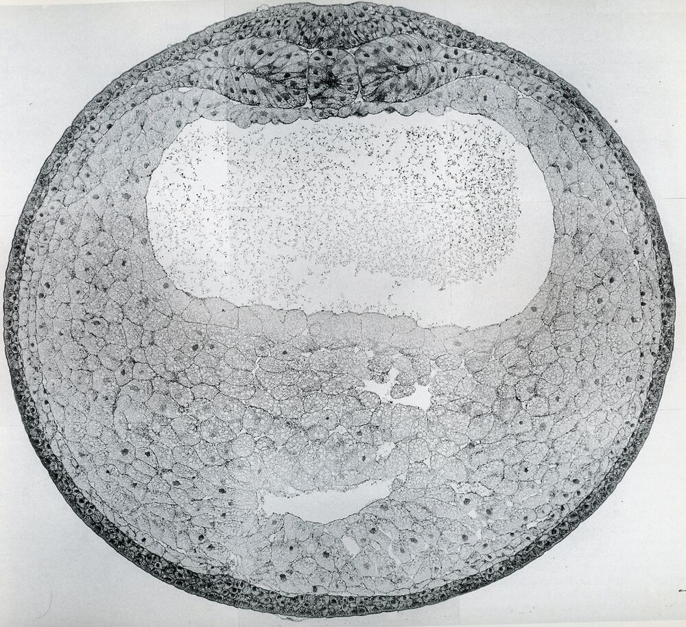
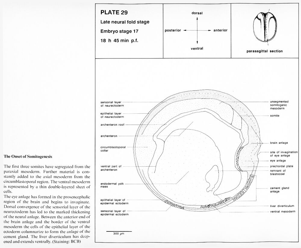
Plate 29: Embryo stage 17, Late neural fold stage. The onset of somitogenesis. 18 h 45 min p.f.
Higher resolution Plate 29.
Higher resolution Plate 29.
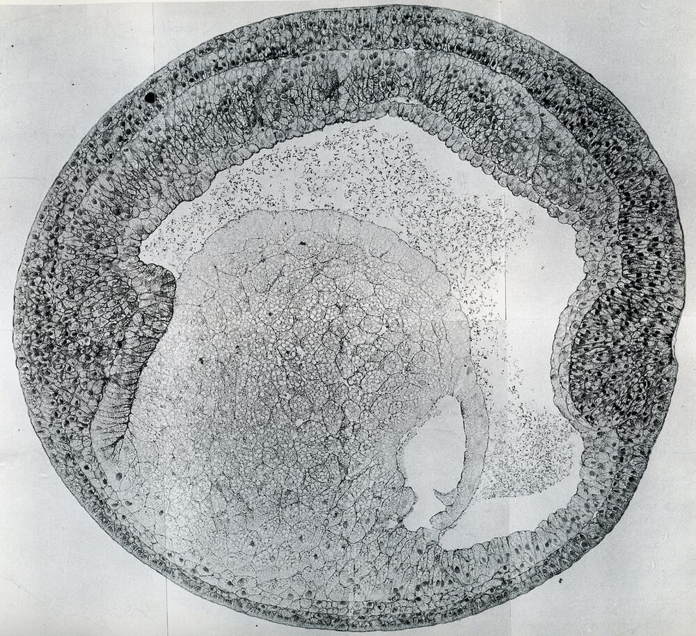
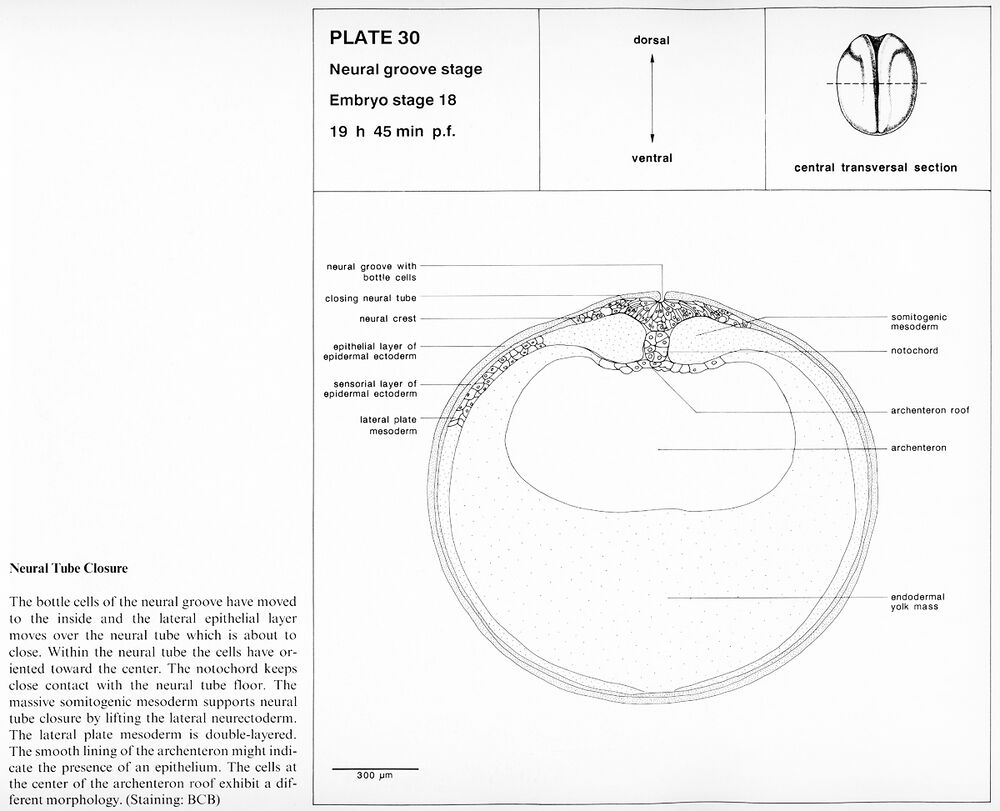
Plate 30: Embryo stage 18, Neural groove stage. Neural tube closure. 19 h 45 min p.f.
Higher resolution Plate 30.
Higher resolution Plate 30.
Neural tube stage
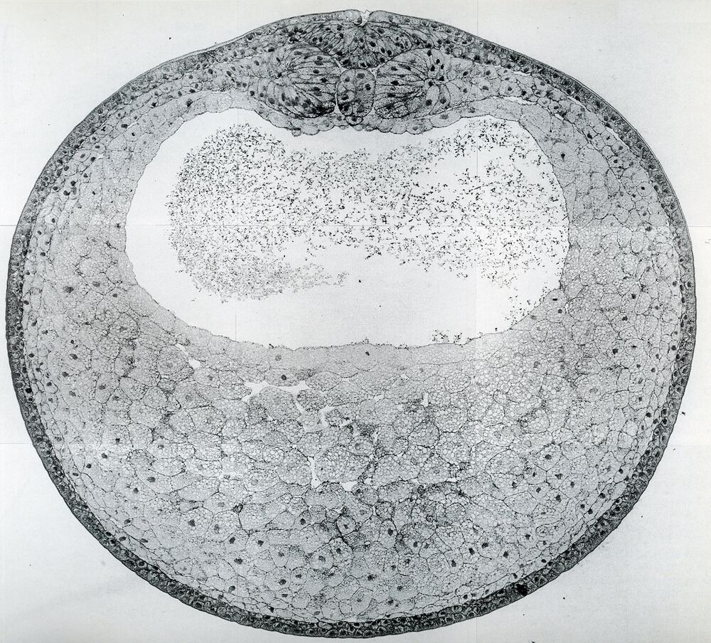
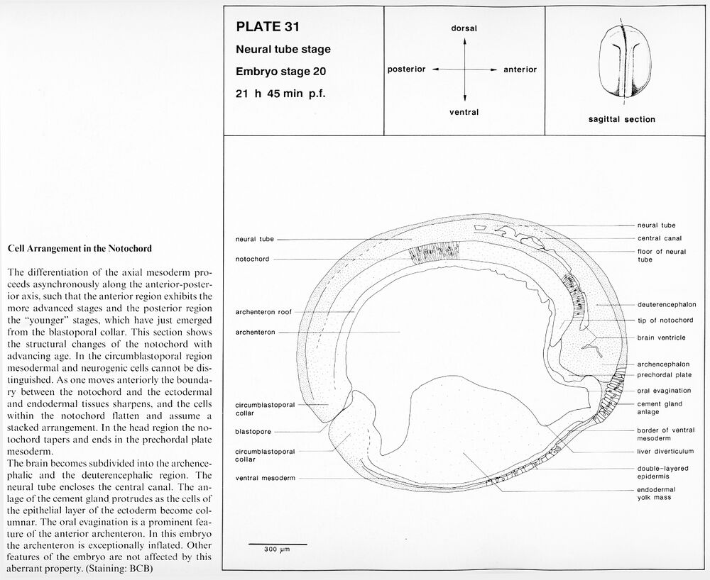
Plate 31: Embryo stage 20, Neural tube stage. Cell arrangement in the notochord. 21 h 45 min p.f.
Higher resolution Plate 31.
Higher resolution Plate 31.
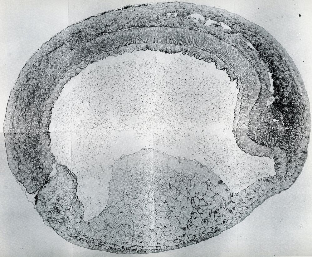
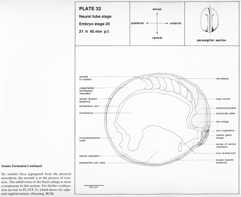
Plate 32: Embryo stage 20, Neural tube stage. Somite formation continued. 21 h 45 min p.f.
Higher resolution Plate 32.
Higher resolution Plate 32.
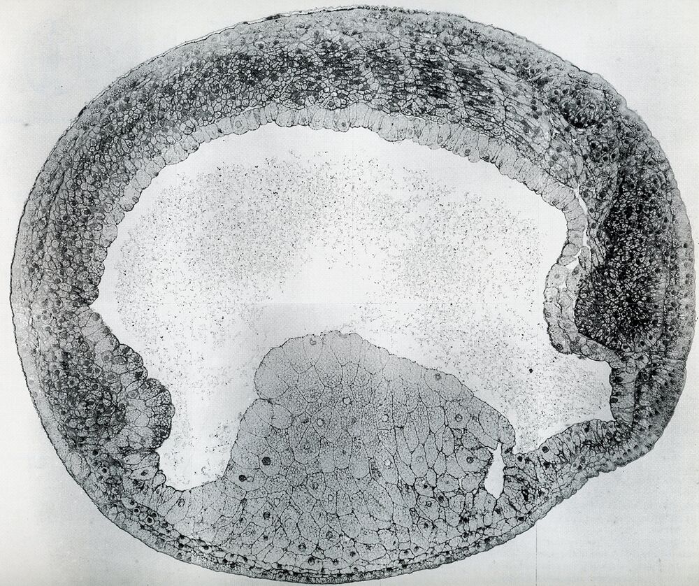
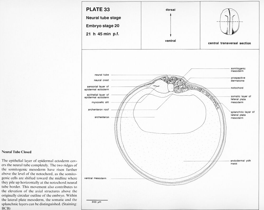
Plate 33: Embryo stage 20, Neural tube stage. Neural tube closed. 21 h 45 min p.f.
Higher resolution Plate 33.
Higher resolution Plate 33.
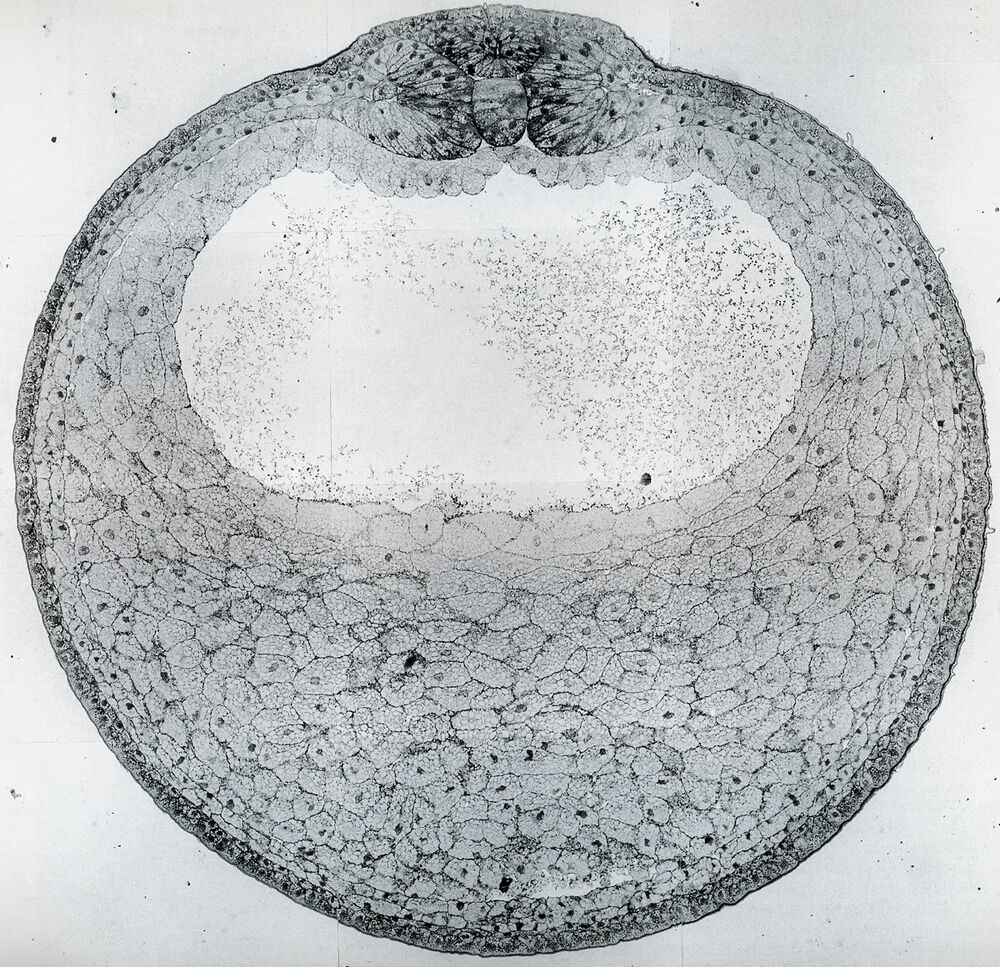
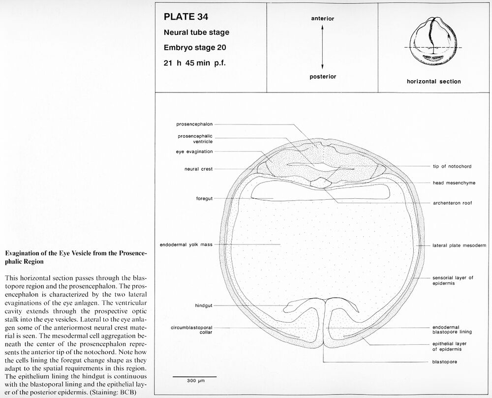
Plate 34: Embryo stage 20, Neural tube stage. Evagination of the eye vesicle from the proencephalic region. 21 h 45 min p.f.
Higher resolution Plate 34.
Higher resolution Plate 34.
Early tail bud stage
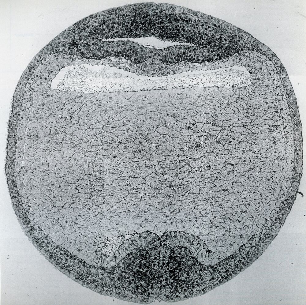
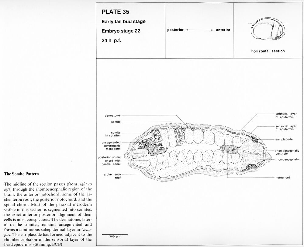
Plate 35: Embryo stage 22, Early tail bud stage. The somite pattern. 24 h p.f.
Higher resolution Plate 35.
Higher resolution Plate 35.
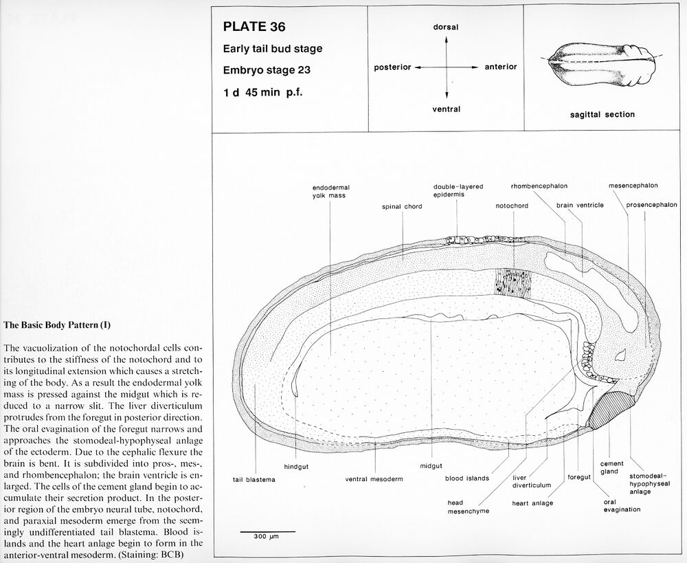
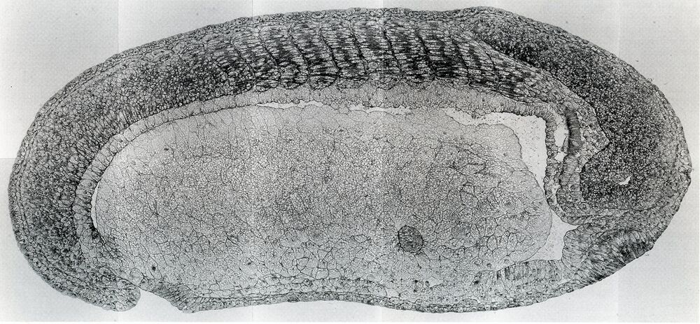
Plate 36: Embryo stage 23, Early tail bud stage. The basic body pattern (I). 1 d 45 min p.f.
Higher resolution Plate 36.
Higher resolution Plate 36.
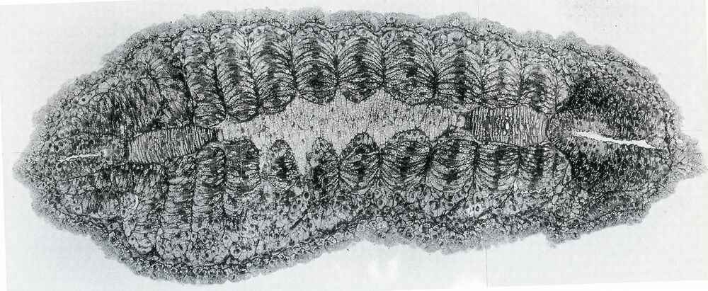
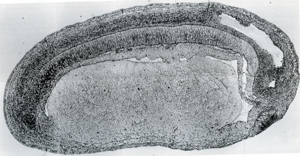
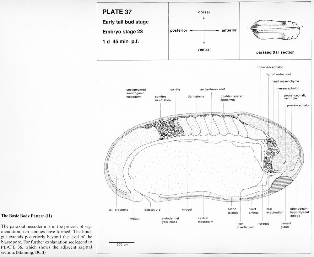
Plate 37: Embryo stage 23, Early tail bud stage. The basic body pattern (II). 1 d 45 min p.f.
Higher resolution Plate 37.
Higher resolution Plate 37.
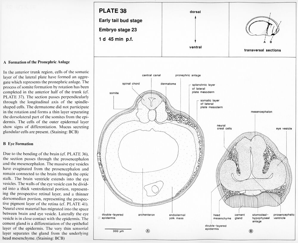
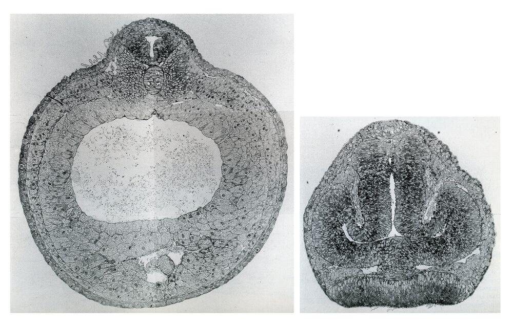
Plate 38: Embryo stage 23, Early tail bud stage. A. Formation of the pronephric anlage. B. Eye formation. 1 d 45 min p.f.
Higher resolution Plate 38a and 38b.
Higher resolution Plate 38a and 38b.
Tail bud stage
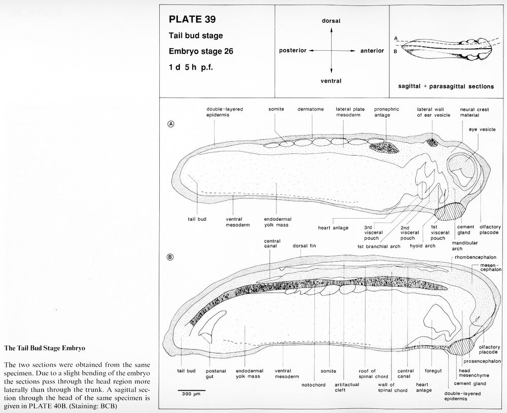
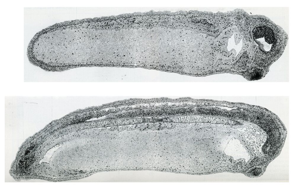
Plate 39: Embryo stage 26, Tail bud stage. The tail bud stage embryo. 1d 5 h p.f.
Higher resolution Plate 39a and 39b.
Higher resolution Plate 39a and 39b.
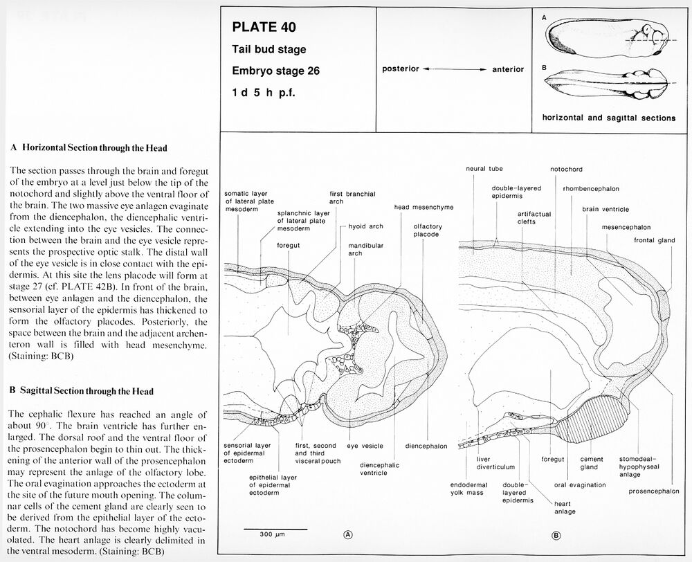
Plate 40: Embryo stage 26, Tail bud stage. A. Horizontal section through the head. B. Sagittal section through the head. 1d 5 h p.f.
Higher resolution Plate 40a and 40b.
Higher resolution Plate 40a and 40b.
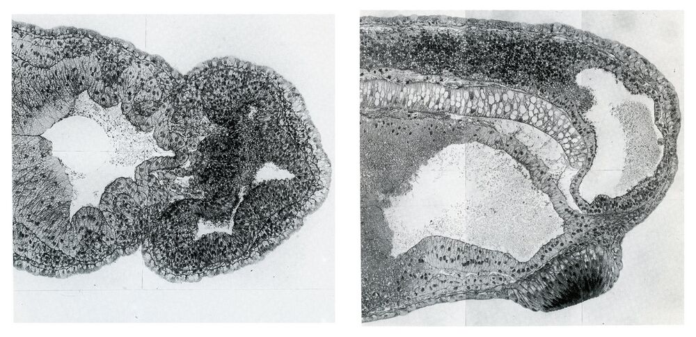
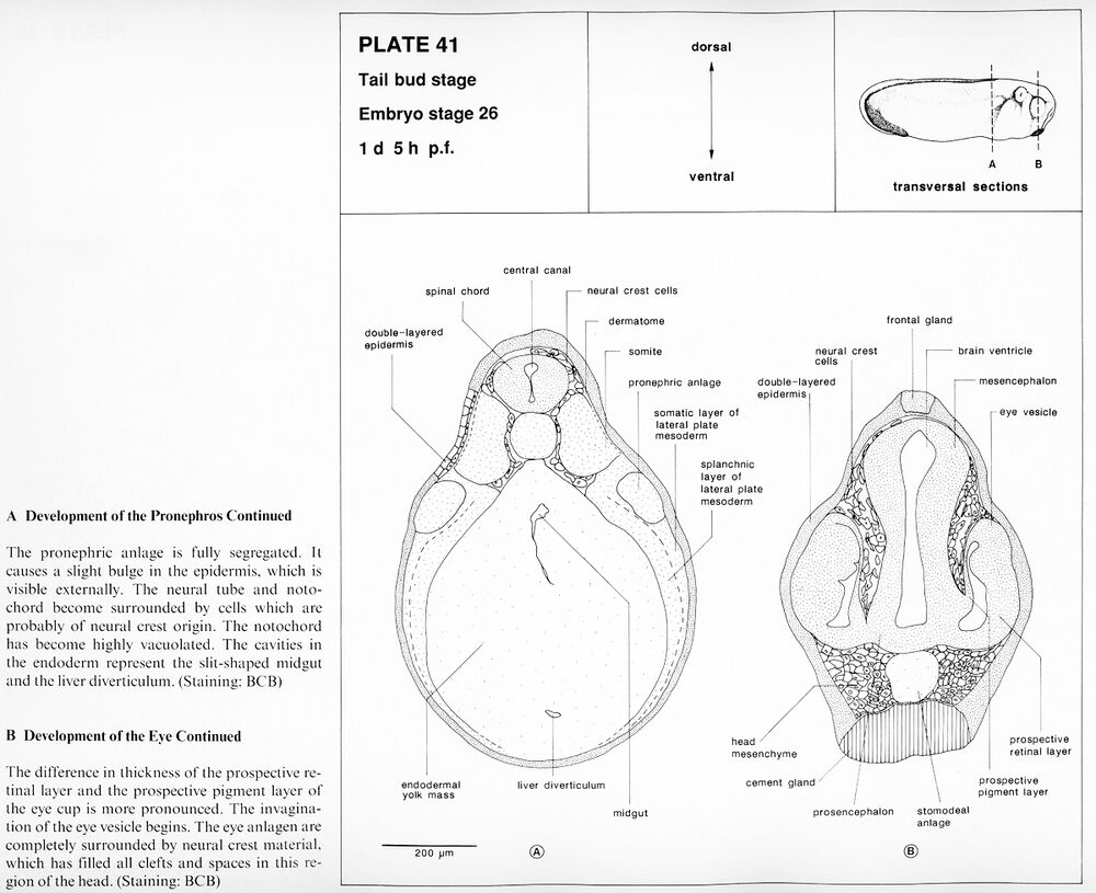
Plate 41: Embryo stage 26, Tail bud stage. A. Development of the pronephros continued. B. Development of the eye continued. 1d 5 h p.f.
Higher resolution Plate 41a and 41b.
Higher resolution Plate 41a and 41b.
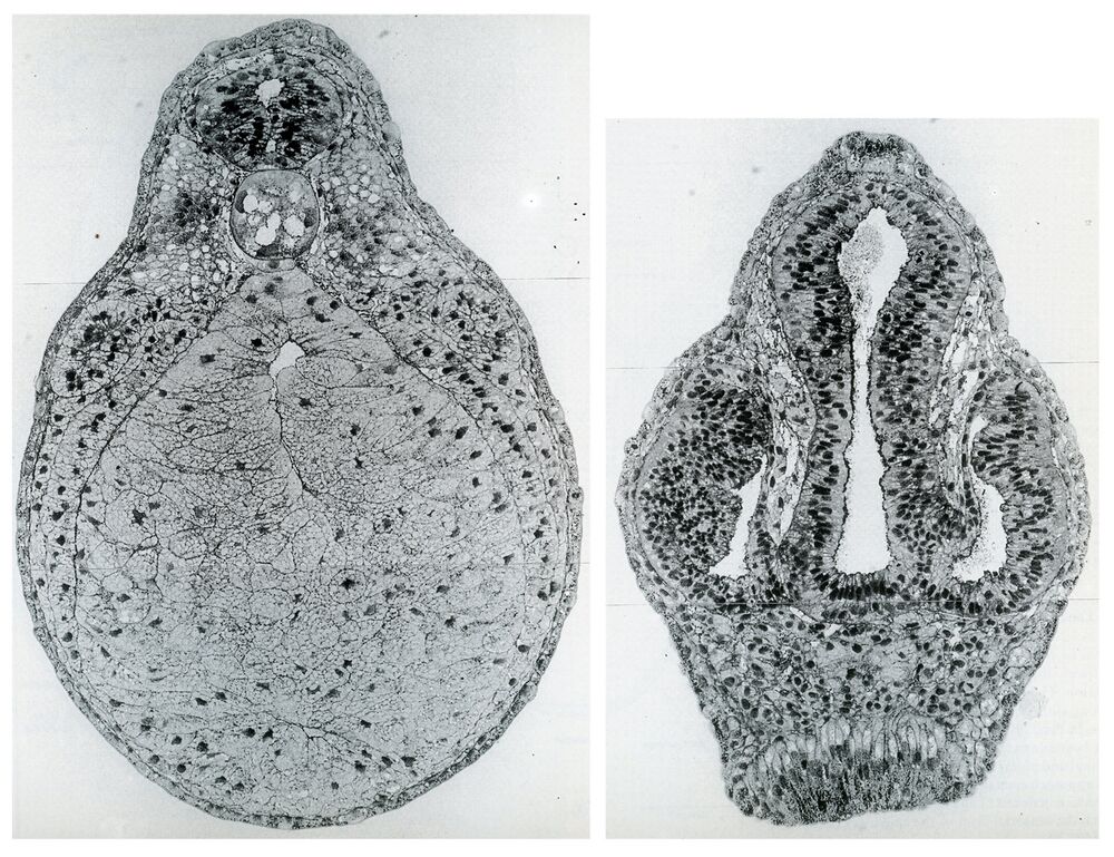
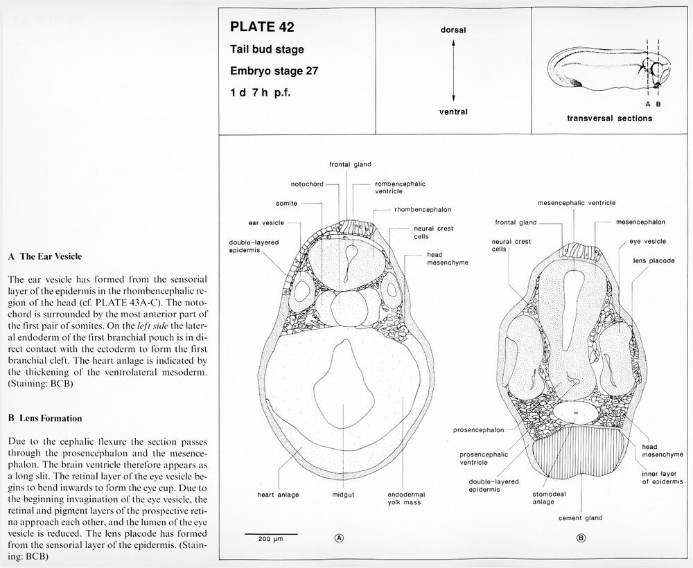
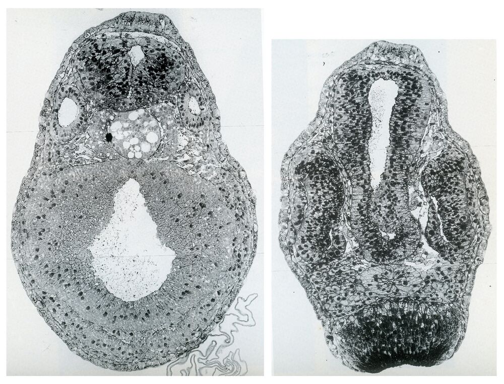
Plate 42: Embryo stage 27, Tail bud stage. A. The ear vesicle. B. Lens formation. 1d 7 h p.f.
Higher resolution Plate 42a and 42b.
Higher resolution Plate 42a and 42b.
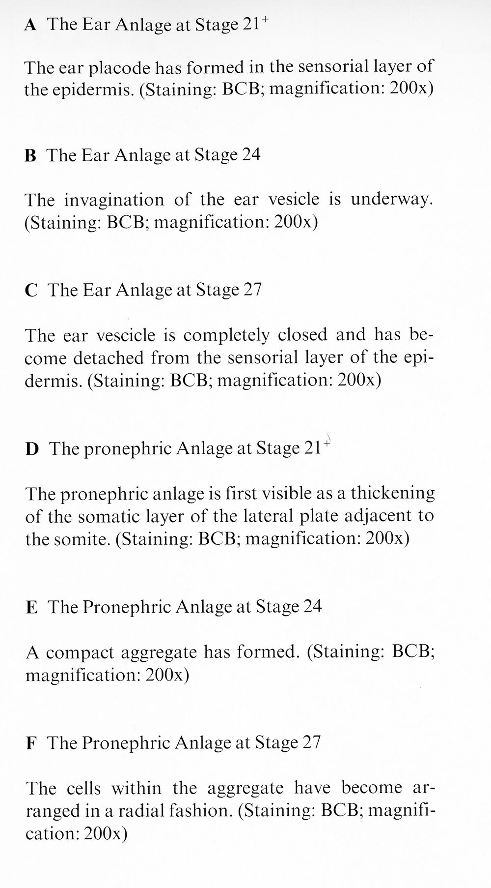
Early stages of ear and
pronephros development
at higher magnifications
pronephros development
at higher magnifications
Last Updated: 2019-09-01

