XB-IMG-134064
Xenbase Image ID: 134064

|
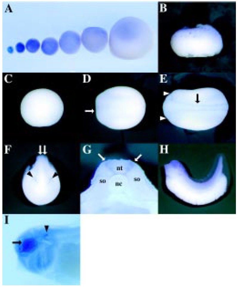
Fig. 5. Spatial expression of Xoom during
oogenesis and embryogenesis. Whole-mount
in situ hybridization with digoxigenin-labeled
Xoom RNA probe was performed in albino oocytes
and embryos. (A) Oocyte stage I to VI (left
to right). Animal pole is up. (B) Lateral view of
early blastula (st. 7). Animal pole is up. (C) Animal
view of gastrula (st. 10). (D) Dorsal view of early neurula (st. 15). Zygotic expression occurs in the dorsoanterior region
(arrow). (E) Dorsal view of late neurula (st. 19). Expression of Xoom is
localized in both sides of the neural tube (arrow) and upper space of the eye
vesicles (arrowheads). (F) Anterior view of late neurula (st. 19). Expression
of Xoom is localized in both sides of neural tube (arrow) and upper space of
eye vesicles (arrowheads). (G) Transverse section at the trunk region of late
neurula (st. 19). nt, neural tube; nc, notochord; so, somite. Xoom specifically
expresses in neural crest cells around the neural tube. (H) Lateral view of
tadpole (st. 35). (I) High magnification of cleared tadpole head. Intense
expression of Xoom is observed in the optic vesicle (arrow) and the otic
vesicle (arrowhead). Image published in: Hasegawa K et al. (1999) Copyright © 1999. Image reproduced with permission of the Publisher.
Image source: Published Larger Image Printer Friendly View |
