XB-IMG-135971
Xenbase Image ID: 135971

|
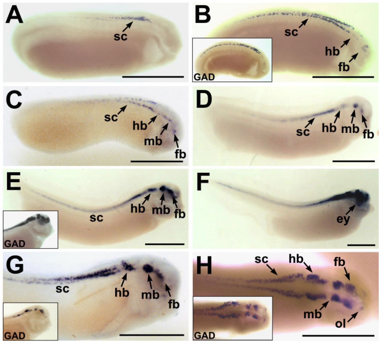
Fig. 3. Whole mount xGAT-1 expression in the developing Xenopus
embryo. Insets show a stage-matched embryo hybridized with an
antisense GAD probe. A: xGAT-1 expression is first detected in the
spinal cord during late neurula stages (stage 21). B: xGAT-1 signal is
present in the hindbrain and forebrain by stage 23. C: By stage 26,
expression increases in the spinal cord and can be observed in all
parts of the developing brain. DâF: xGAT-1 expression continues to
strengthen during tailbud stages (D, stage 28; E, stage 32), and by
swimming tadpole stages (F, stage 40) is present at high levels
throughout the central nervous system and eye. G: Higher magnification
of the head of a stage 33 embryo showing distinct domains of
xGAT-1 expression in the forebrain, midbrain, and hindbrain. H: Dorsal
view of a tailbud stage embryo (stage 32). fb, forebrain; hb, hindbrain;
mb, midbrain; ol, olfactory placodes; sc, spinal cord. Scale bar
1 mm in AâH. Image published in: Li M et al. (2006) Copyright © 2006. Image reproduced with permission of the Publisher.
Image source: Published Larger Image Printer Friendly View |
