XB-IMG-154155
Xenbase Image ID: 154155

|
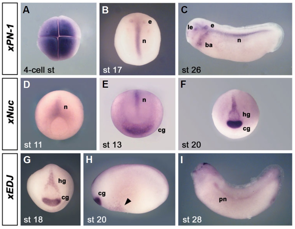
Fig. 6. Whole-mount in situ hybridization of xPN-1, xNuc and xEDJ. Embryos are shown in animal
(A), dorsal (B,D), lateral (C,H,I) or anterior view (E-G). (A-C) Xenopus Proteinase Nexin-1 (xPN-1). (A)
Four-cell stage embryo showing high level of maternal transcripts. (B) Late neurula. Note expression
in the notochord (n) and ear placode (e). (C) Early tail bud stage with additional expression domains in
the lens (le) and ectoderm of the branchial arches (ba). (D-F) Xenopus Nucleobindin (xNuc). (D) Midgastrula
showing expression in the notochord (n). (E) Early neurula with additional signal in the cement
gland (cg). (F) Late neurula. Note strong expression in the cement gland and hatching gland (hg). (G-I)
Xenopus ER-associated DNAJ chaperone (xEDJ). (G) Late neurula with expression in cement gland (cg)
and hatching gland (hg). (H) Late neurula depicting faint signal on the ventral side (arrowhead). (I) Early
tail bud stage. Note expression in the pronephros (pn) and in the hatching gland in the dorsal head. Image published in: Pera EM et al. (2005) Copyright © 2005. Image reproduced with permission of the Publisher, University of the Basque Country Press.
Image source: Published Larger Image Printer Friendly View |
