XB-IMG-44177
Xenbase Image ID: 44177
|
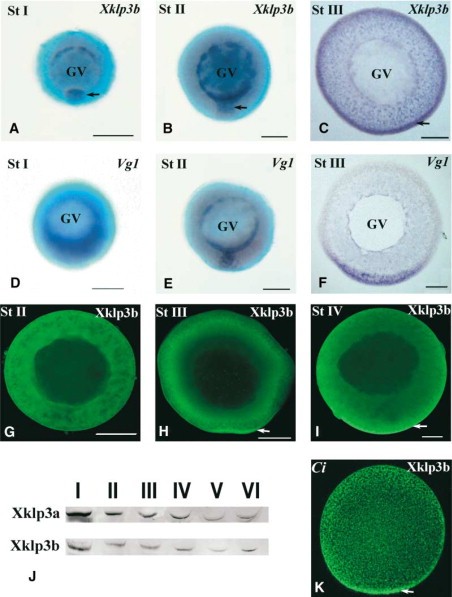
Figure 1. Expression and Localization of Heterotrimeric Kinesin II mRNA and Protein during Oogenesis(AâF) Whole mount in situ hybridization to endogenous Xklp3b (AâC) and Vg1 mRNA (DâF) in stage I (A and D), stage II (B and E), and stage III (C and F) oocytes. Arrows in (A)â(C) depict slight enrichment of Xklp3b mRNA in the mitochondrial cloud, wedge region, and vegetal cortex, respectively. GV is the germinal vesicle. The vegetal pole is oriented down in all images, and (C) and (F) are sectioned so that the cortex is visible. (GâI and K) Confocal images of Xklp3b-labeled Xenopus stage II (G), stage III (H), and stage IV (I) oocytes and an egg from Ciona intestinalis (K). Arrows in (H), (I), and (K) indicate enrichment of kinesin II at the vegetal cortex. (J) Western blots of whole-cell extracts prepared from stage IâVI oocytes via Xklp3a- and Xklp3b-specific antibodies. The scale bar represents 100 μm. Image published in: Betley JN et al. (2004) Copyright © 2004. Image reproduced with permission of the Publisher, Elsevier B. V.
Image source: Published Larger Image Printer Friendly View |