XB-IMG-76194
Xenbase Image ID: 76194
|
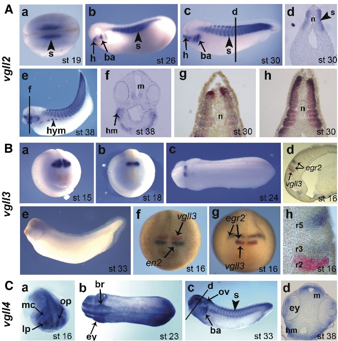
Fig. 3. Spatial expression analysis of Xenopus laevis vestigial like genes in embryo by in situ hybridization. (A) vgll2 expression. (a) dorsal view; (b,c,e) lateral views; (d,f,g,h) transversal sections. Double in situ hybridization of stage 30 embryo with vgll2 and myod1 (g) or vgll2 and myl1 (h). (B) vgll3 expression. (a,b,f,g), frontal views; (c) dorsal view; (e) lateral view. (f) double in situ hybridization of stage 16 embryo with vgll3 and en2 (f) or vgll3 and egr2 (g). (d) parasagittal section of the embryo shown in g; (h) higher magnification of d. The position of rhombomeres is indicated (r2, r3 and r5). (C) vgll4 expression. (a) anterior view; (b) dorsal view; (c) lateral view; (d) transversal section. The positions of the section were indicated. Embryo staging according to Nieuwkoop and Faber (1967) is indicated. ba, branchial arches; br, brain; ey, eye; h, hypophyseal anlage; hm, head mesenchyme; hym, hypaxial muscle; lp, lens placode; m, mesencephalon; mc, mandibular neural crest; n, notochord; op, olfactory placode; ov, otic vesicle; r, rhombomere; s, somites. Image published in: Faucheux C et al. (2010) Copyright © 2010. Image reproduced with permission of the Publisher, University of the Basque Country Press.
Image source: Published Larger Image Printer Friendly View |