XB-IMG-84316
Xenbase Image ID: 84316
|
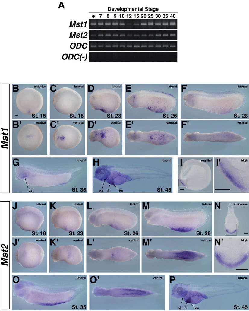
Fig. 1. Expression pattern of Mst1 and Mst2 in Xenopus tropicalis
development. (A) RT-PCR analysis revealed the temporal
expression patterns of Mst1 and Mst2. RNA was extracted from
Xenopus tropicalis embryos at the stages indicated above each
lane. ODC were used as internal controls. ODC(-), RT-PCR without
reverse transcriptase in ODC reaction. e, unfertilized egg. (B-P)
Whole-mount in situ hybridization analysis of Mst1 and Mst2.
(B-H) Localized expression pattern of Mst1. (B�) The anterior view
corresponding to (B). (C�-F�) The ventral view corresponding to
C-F. (I) Hemi-sectioning of a stage-18 embryo revealed detailed
localization of Mst1 in the aVBI. (I�) High magnification view of
the boxed region in (I). (J-M,O,P) Localized expression pattern of
Mst2. (J�-M�,O�) The ventral view corresponding (J-M, P,O). (N)
Transverse sectioning of a stage-28 embryo revealed detailed
localization of Mst2 in the pVBI. (N�) High magnification view
of the boxed region in (N). Scale bars, 100 +m. View direction is
indicated in the upper right of each panel. Developmental stage
is indicated in the lower right. Abbreviations: ba, branchial arch;
du, duodenum; in, intestine. Image published in: Nejigane S et al. (2013) Copyright © 2013. Image reproduced with permission of the Publisher, University of the Basque Country Press.
Image source: Published Larger Image Printer Friendly View |