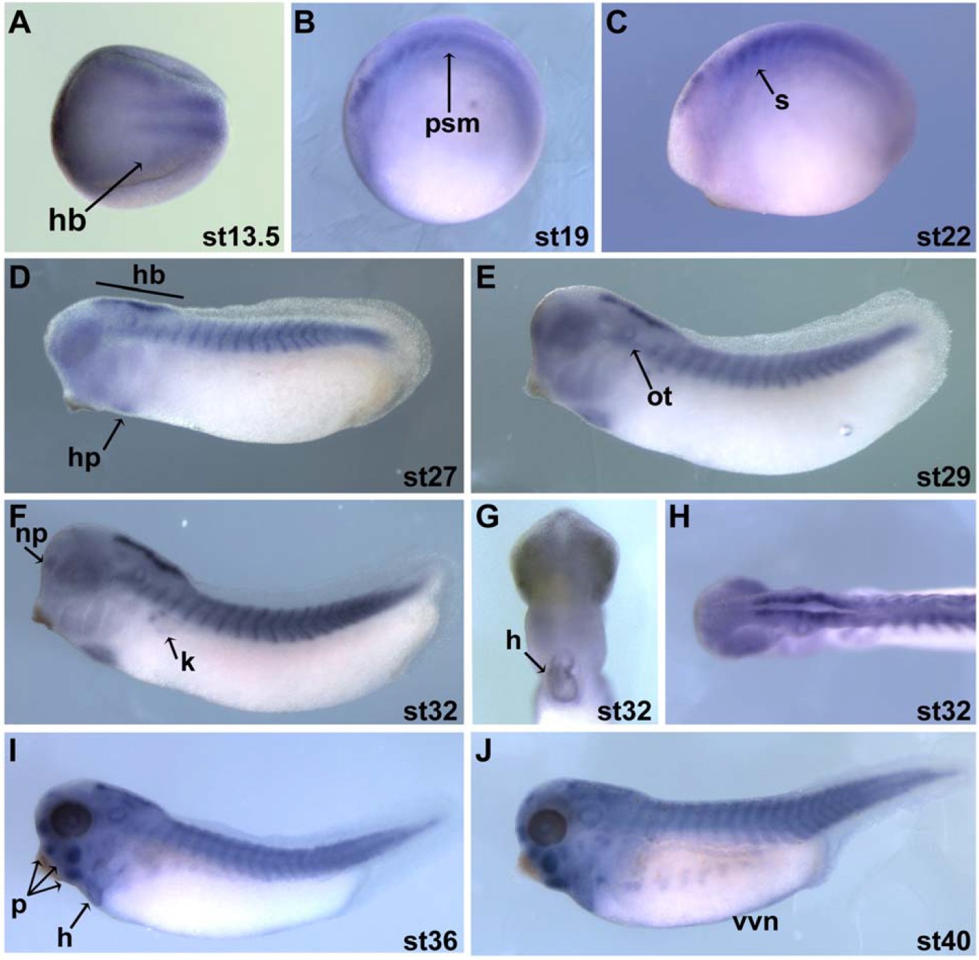
Figure S3. Xenopus tropicalis Cst Spatial Expression Whole mount in situ hybridization of Cst in X. tropicalis of Stage 14 (neurula) to Stage 40 (tadpole) embryos using a Cst-specific probe common to both Cstα and Cstβ. A is an anterior view. B-F, I-J are lateral views with anterior to the left. G is a ventral view with anterior to the top. H is a dorsal view with anterior to the left. hindbrain (hb), presomitic mesoderm (psm), somites (s), heart primordium (hp), otic vesicle (ot), nasal placode (np), facial placodes (p), heart (h), kidney (k), vascular vitelline network (vvn).
Image published in: Christine KS and Conlon FL (2008)
Copyright © 2008. Image reproduced with permission of the Publisher, Elsevier B. V.
| Gene | Synonyms | Species | Stage(s) | Tissue |
|---|---|---|---|---|
| casz1.L | castor, castor-alpha, csta | X. laevis | Sometime during NF stage 13 to NF stage 19 | presomitic mesoderm |
| casz1.L | castor, castor-alpha, csta | X. laevis | Throughout NF stage 22 | somite |
| casz1.L | castor, castor-alpha, csta | X. laevis | Sometime during NF stage 27 to NF stage 29 and 30 | hindbrain somite heart primordium heart |
| casz1.L | castor, castor-alpha, csta | X. laevis | Throughout NF stage 32 | hindbrain pronephric nephrostome heart |
| casz1.L | castor, castor-alpha, csta | X. laevis | Sometime during NF stage 35 and 36 to NF stage 40 | hindbrain somite heart musculature of face hypaxial muscle |
Image source: Published
Permanent Image Page
Printer Friendly View
XB-IMG-130275