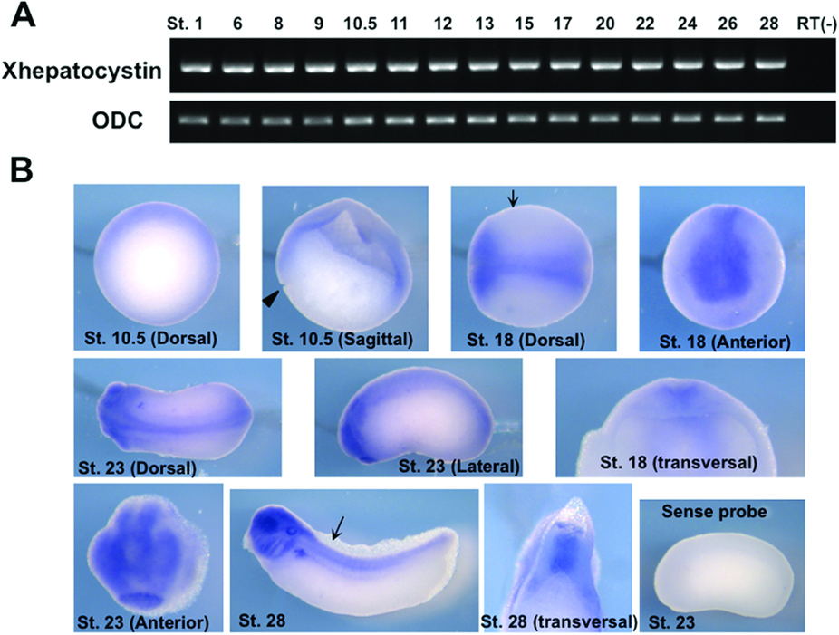
Figure 2: Expression pattern of Xhepatocystin during early development. (A) Xhepatocystin is expressed throughout embryogenesis as monitored by RT-PCR analysis. Ornithine decarboxylase (ODC) was used as a loading control. (B) Expression pattern of Xhepatocystin at selected developmental stages as analyzed by whole-mount in situ hybridization using a Xhepatocystin anti-sense probe. A sense probe was used as a negative control. Arrows indicate the region where the embryo was vertically sectioned and examined. ST: stage. Xhepatocystin was expressed in dorsal ectoderm and dorsal mesoderm. In neurula stage embryos (stage 18 & 23) Xhepatocystin was highly expressed in the neural plate and anterior neural fold. At the early tadpole stage, Xhepatocystin was found expressed in the head, neural tube and notochord. Later in development, strong expression was observed in head, eyes, spinal cord, notochord, liver and kidney.
Image published in: Overton JD et al. (2015)
Copyright © 2015, Macmillan Publishers Limited. Creative Commons Attribution license
| Gene | Synonyms | Species | Stage(s) | Tissue |
|---|---|---|---|---|
| prkcsh.L | hepatocystin | X. laevis | Throughout NF stage 10.5 | ectoderm mesoderm dorsal |
| prkcsh.L | hepatocystin | X. laevis | Throughout NF stage 18 | neural plate pre-chordal neural plate chordal neural plate neural crest cranial neural crest |
| prkcsh.L | hepatocystin | X. laevis | Throughout NF stage 23 | neural tube posterior neural tube anterior neural tube cement gland primordium optic vesicle migratory neural crest cell |
| prkcsh.L | hepatocystin | X. laevis | Sometime during NF stage 28 to NF stage 35 and 36 | pharyngeal arch mandibular arch hyoid arch branchial arch pronephric kidney pronephric tubule pronephric duct notochord central nervous system brain spinal cord eye forebrain midbrain hindbrain otic vesicle |
Image source: Published
Permanent Image Page
Printer Friendly View
XB-IMG-148631