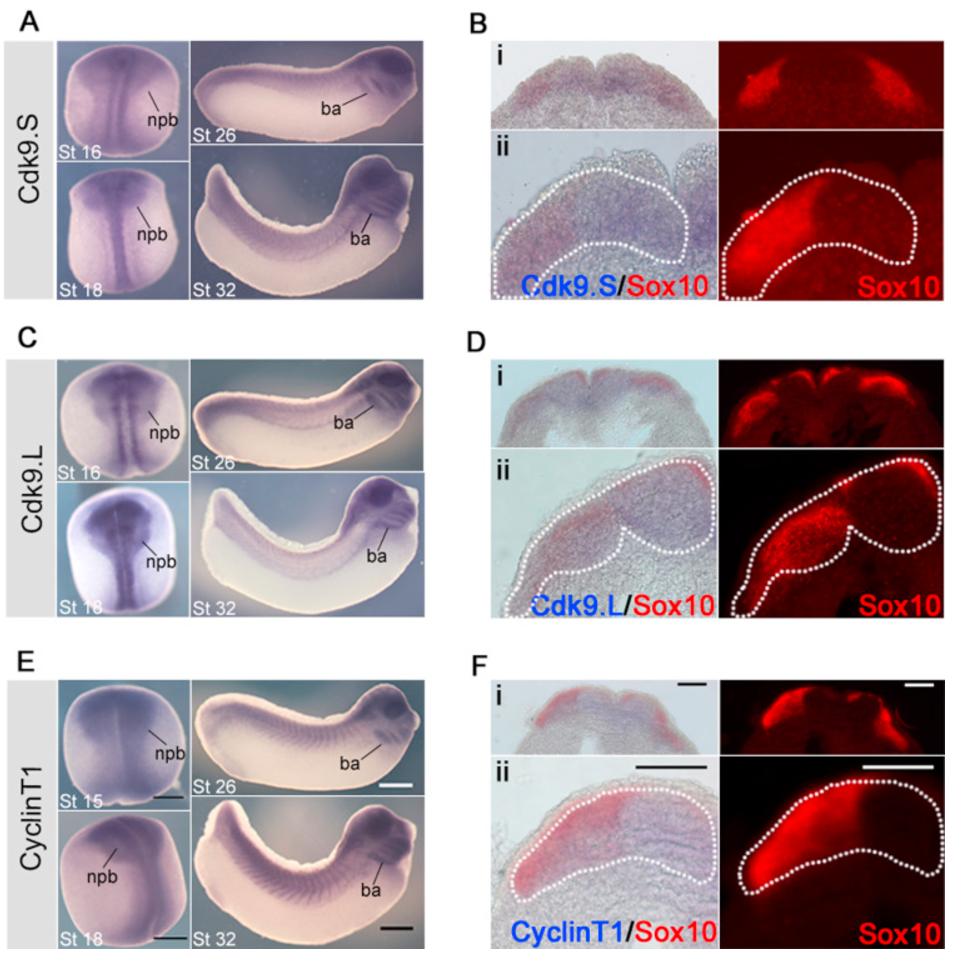
Fig. 1. The expression of the P-TEFb complex components during Xenopus laevis development. Transcripts for the P-TEFb components were detected using NBT/BCIP and expression of CDK9.S, CDK9.L and CyclinT1 appeared blue in the sectioned neurula stage embryo. Sox10 was detected using fast red and fluorescence microscopy. (A) CDK9.S expression at stage 16 and 18 in the neural plate and neural plate border and expression at stages 26 and 32 in the branchial arches. Scale bar=100 µm (Bi) 10à magnification section of a stage 16 embryo showing CDK9.S expression in the neural plate and neural plate border (blue) and Sox10 expression in the neural crest (red). (ii) 20à magnification section shows overlap of the blue CDK9.S expression with the red Sox10 expression. Expression of CDK9.S is outlined with a dashed white line and overlaid onto the Sox10 expression image. Scale bar=100 µm (C) CDK9.L expression at stage 16 and 18 in the neural plate and neural plate border and expression at stages 26 and 32 in the branchial arches. Scale bar=100 µm (Di) 10à magnification section of a stage 16 embryo showing CDK9.L expression in the neural plate and neural plate border (ii) 20à magnification section shows overlap of the blue CDK9.L expression with the red Sox10 expression. Expression of CDK9.L is outlined with a dashed white line and overlaid onto the Sox10 expression image. Scale bar=100 µm (E) CyclinT1 expression at stage 15 and 18 in the neural plate and neural plate border and expression at stages 26 and 32 in the branchial arches. Scale bar=100 µm (Fi) 10à magnification section of a stage 15 embryo showing CDK9.S expression in the neural plate and neural plate border (blue) and Sox10 expression in the neural crest (red). (ii) 20à magnification section shows overlap of the blue CDK9.S expression with the red Sox10 expression. Expression of CDK9.S is outlined with a dashed white line and overlaid onto the Sox10 expression image. Scale bar=100 µm. Npb=Neural plate border, ba=branchial arches.
Image published in: Hatch VL et al. (2016)
Copyright © 2016. Image reproduced with permission of the Publisher, Elsevier B. V.
| Gene | Synonyms | Species | Stage(s) | Tissue |
|---|---|---|---|---|
| ccnt1.L | X. laevis | Throughout NF stage 15 | neural plate neural plate border neural crest | |
| sox10.L | X. laevis | Sometime during NF stage 15 to NF stage 16 | neural plate border neural crest | |
| cdk9.S | c-2k, cdc2l4, cdk9-a, cdk9-b, ctk1, pitalre, tak | X. laevis | Throughout NF stage 16 | neural plate neural plate border neural crest |
| cdk9.L | c-2k, cdc2l4, cdk9-a, cdk9-b, ctk1, pitalre, tak | X. laevis | Throughout NF stage 16 | neural plate neural plate border neural crest |
| cdk9.S | c-2k, cdc2l4, cdk9-a, cdk9-b, ctk1, pitalre, tak | X. laevis | Throughout NF stage 18 | neural plate neural plate border neural crest |
| cdk9.L | c-2k, cdc2l4, cdk9-a, cdk9-b, ctk1, pitalre, tak | X. laevis | Throughout NF stage 18 | neural plate neural plate border neural crest |
| ccnt1.L | X. laevis | Throughout NF stage 18 | neural plate neural plate border neural crest | |
| cdk9.S | c-2k, cdc2l4, cdk9-a, cdk9-b, ctk1, pitalre, tak | X. laevis | Throughout NF stage 26 | pharyngeal arch head brain |
| cdk9.L | c-2k, cdc2l4, cdk9-a, cdk9-b, ctk1, pitalre, tak | X. laevis | Throughout NF stage 26 | pharyngeal arch head brain |
| ccnt1.L | X. laevis | Throughout NF stage 26 | pharyngeal arch brain | |
| cdk9.L | c-2k, cdc2l4, cdk9-a, cdk9-b, ctk1, pitalre, tak | X. laevis | Throughout NF stage 28 | branchial arch head brain |
| cdk9.S | c-2k, cdc2l4, cdk9-a, cdk9-b, ctk1, pitalre, tak | X. laevis | Throughout NF stage 32 | branchial arch head brain |
| ccnt1.L | X. laevis | Throughout NF stage 32 | branchial arch brain |
Image source: Published
Permanent Image Page
Printer Friendly View
XB-IMG-154127