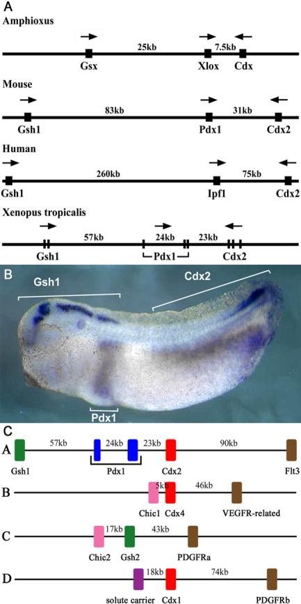
Figure 1. The Xenopus tropicalis ParaHox cluster. A: Diagram of ParaHox clusters from amphioxus, mouse, human, and X. tropicalis (not to scale). Arrows denote the transcription orientation of the genes. The X. tropicalis cluster diagram indicates exon/intron structure. B: One-colour triple WISH on a stage 30 X. tropicalis embryo with probes for Xt Gsh1, Xt Pdx1, and Xt Cdx2. C: The remnants of the four X. tropicalis ParaHox clusters, showing some of the flanking genes and approximate distances. Gsx genes are shown in green, Xlox in blue, Cdx in red, Chic in pink, and tyrosine kinase receptors in brown.
Image published in: Illes JC et al. (2009)
Copyright © 2009. Image reproduced with permission of the Publisher, John Wiley & Sons.
| Gene | Synonyms | Species | Stage(s) | Tissue |
|---|---|---|---|---|
| cdx2 | cad2, cdx-2, cdx3-like, xcad2, Xcaudal2 | X. tropicalis | Throughout NF stage 29 and 30 | endoderm spinal cord posterior neural tube hindgut tail region dorsal tail fin |
| gsx1 | gsh1, Xgsh1 | X. tropicalis | Throughout NF stage 29 and 30 | brain forebrain midbrain hindbrain rhombomere hypothalamus |
| pdx1 | ipf1, pdx-1, STF-1, XlHbox-8, XlHbox8 | X. tropicalis | Throughout NF stage 29 and 30 | endoderm pancreas foregut |
Image source: Published
Permanent Image Page
Printer Friendly View
XB-IMG-26181