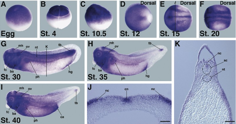
Fig. 5. Spatial expression pattern of xtYap during the early develop- ment of Xenopus tropicalis. (A-K) Spatial expression of xtYap was ana- lyzed by whole-mount in situ hybrid- ization. Anterior is at the left of each panel. (A) Unfertilized egg, (B) 8-cell stage, (C) early gastrula, (G) tailbud stage, (H) late tailbud stage, and (I) tadpole stage in lateral view. (D) Late gastrula, (E) neurula stage, and (F) late neurula stage in dorsal view. (J) Transverse section of (E). (K) Trans- verse section of (G). The line position is indicated in (E,G). Scale bars, 50 ïm. Abbreviations: ba, branchial arch; ca, cloaca; cn, center of neural plate; fc, facial connective tissues; hg, hind- gut; mh, midbrain-hindbrain boundary; nc, neural crest; nt, notochord; ov, otic vesicle; ph, pronephros; sc, spinal cord; tb, tailbud.
Image published in: Nejigane S et al. (2011)
Copyright © 2011. Image reproduced with permission of the Publisher, University of the Basque Country Press.
| Gene | Synonyms | Species | Stage(s) | Tissue |
|---|---|---|---|---|
| yap1 | xyap, yap, yap2, yap65, yki | X. tropicalis | Throughout NF stage 10.5 | animal cap |
| yap1 | xyap, yap, yap2, yap65, yki | X. tropicalis | Throughout NF stage 12 | involuted dorsal mesoderm |
| yap1 | xyap, yap, yap2, yap65, yki | X. tropicalis | Throughout NF stage 15 | chordal neural plate cranial neural crest |
| yap1 | xyap, yap, yap2, yap65, yki | X. tropicalis | Throughout NF stage 20 | intermediate mesoderm neural tube cranial neural crest |
| yap1 | xyap, yap, yap2, yap65, yki | X. tropicalis | Throughout NF stage 29 and 30 | otic vesicle midbrain-hindbrain boundary notochord hindgut trunk neural crest tail bud spinal cord pronephric nephron late proximal tubule late distal tubule pronephric tubule connective tissue head region pharyngeal arch branchial arch post-anal gut |
| yap1 | xyap, yap, yap2, yap65, yki | X. tropicalis | Throughout NF stage 35 and 36 | otic vesicle midbrain hindbrain midbrain-hindbrain boundary hindgut eye connective tissue head region pharyngeal arch branchial arch pronephric nephrostome pronephric nephron late proximal tubule late distal tubule pronephric tubule dorsal longitudinal anastomosing vessel |
| yap1 | xyap, yap, yap2, yap65, yki | X. tropicalis | Throughout NF stage 40 | otic vesicle midbrain hindbrain midbrain-hindbrain boundary tail bud pronephric kidney connective tissue head region pharyngeal arch branchial arch cloaca |
| yap1 | xyap, yap, yap2, yap65, yki | X. tropicalis | Throughout NF stage 4 (8-cell) | animal blastomere animal |
| yap1 | xyap, yap, yap2, yap65, yki | X. tropicalis | Throughout unfertilized egg stage | animal pole |
Image source: Published
Permanent Image Page
Printer Friendly View
XB-IMG-75395